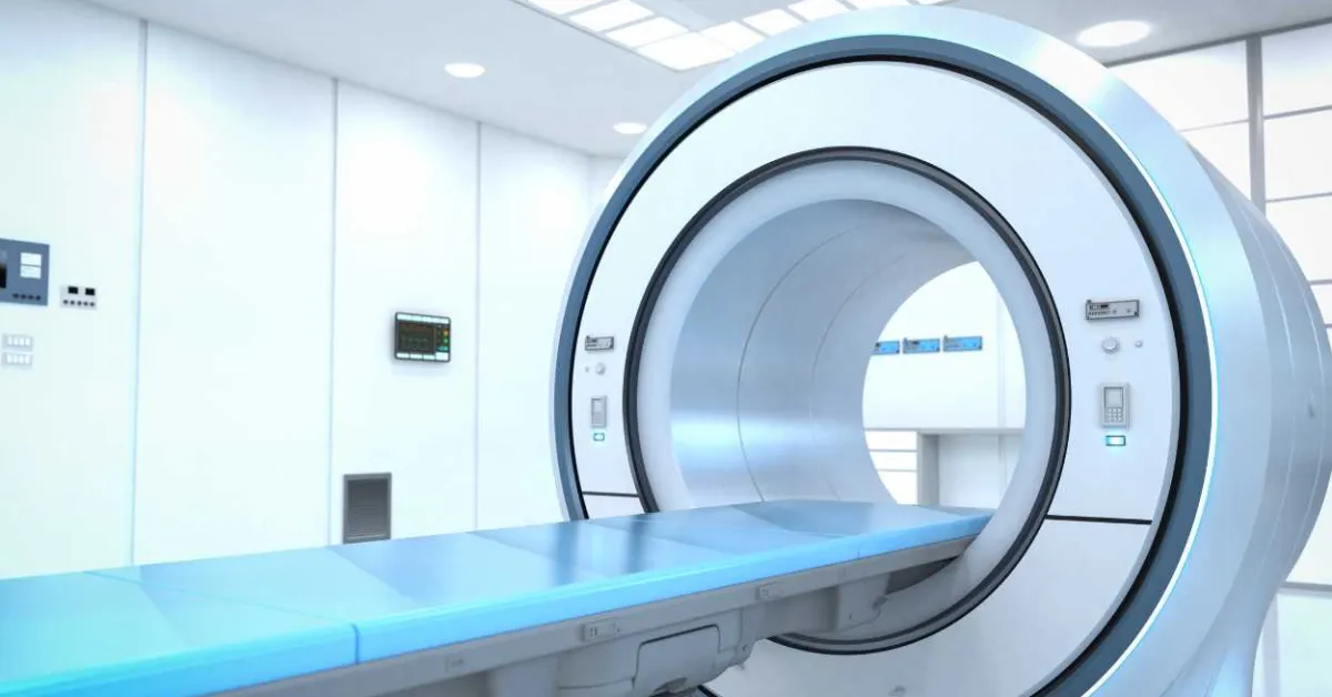
Researchers at the University of Illinois Urbana-Champaign have developed an innovative magnetic resonance imaging (MRI) method capable of noninvasively mapping metabolic activity in the brain, offering new insights into neurological diseases. The technique, called functional glucose MRI (fGluMRI), measures how different regions of the brain consume glucose, a key energy source.
Unlike traditional fMRI, which tracks blood flow as an indirect marker of neural activity, fGluMRI directly monitors metabolic processes by using a safe, non-radioactive glucose analog and novel MRI acquisition methods. This allows researchers to observe real-time metabolic changes with greater spatial and temporal resolution.
In a proof-of-concept study published in Nature Biomedical Engineering, the team demonstrated the technique in both animal models and human participants. The method revealed distinct patterns of glucose utilization associated with various brain functions and potential disease states, including Alzheimer’s disease and cancer.
One of the key advantages of fGluMRI is that it does not rely on radioactive tracers like those used in PET scans, making it safer and more accessible for broader clinical and research use.
This metabolic mapping capability opens up new possibilities for early detection and characterization of neurological disorders by identifying specific “metabolic fingerprints.” The researchers anticipate that fGluMRI could eventually help clinicians monitor disease progression, evaluate treatment response, and improve diagnosis of conditions that alter brain metabolism. As further clinical validation continues, this technology could become a powerful tool in precision medicine and neuroscience.