{ DOWNLOAD AS PDF }
ABOUT AUTHORS
SUJATHA RAMASAMY 1*, SRIKANTA PRIYADARSHAN PATRA 1,PULAK RANJAN BASAK 1,
1Technology Information, Forecasting and Assessment Council (TIFAC)-DST, New Delhi, India.
*sujathar23@gmail.com
ABSTRACT:
Breast cancer is one of the leading type of cancer in India. Current methods used in detection of cancer like mammography combined with Digital Infrared Imaging can help learn about earlier stages of cancer with accuracy.The Niramai an Indian startup developed a software with machine learning and artificial intelligence which can detect and sense cancer cells that are 5 times smaller than what has been done presently. The method is non-contact, painless and free of radiation. This method can help detection of cancer in very early stage before showing any suspicious or alarming signs.
[adsense:336x280:8701650588]
Reference Id: PHARMATUTOR-ART-2592
|
PharmaTutor (Print-ISSN: 2394 - 6679; e-ISSN: 2347 - 7881) Volume 6, Issue 7 Received On: 07/05/2018; Accepted On: 24/05/2018; Published On: 01/07/2018 How to cite this article: Ramasamy, S., Patra, S. and Basak, P. 2018. Advanced technology on accuracy in detection of breast cancer. PharmaTutor. 6, 7 (Jun. 2018), 1-4. DOI:https://doi.org/10.29161/PT.v6.i7.2018.1. |
INTRODUCTION:
The leading type of cancer in women is Breast Cancer. According to data from SEER Cancer Statistics Review report one in every 8 women in US is at the risk of developing a breast abnormality in her lifetime (cancer.gov). It is a well known fact that early detection is very critical in saving a life of a cancer patient. Thermography, also called thermal imaging, uses a special camera to measure the temperature of the skin on the breast's surface for cancer detection. The method involves no radiation and non-invasive test.
Digital Infrared Imaging (DII) which senses metabolic activity and vascular circulation in pre-cancerous tissues and surrounding area. The emitted heat from the body gets captured in the form of images of different variation. Ultra-sensitive medical infrared cameras and sophisticated computers are used to detect, analyze, and produce high-resolution images of these temperature variations. The method is extremely sensitive and the temperature variations may be among the earliest signs of breast cancer and/or a pre-cancerous state of the breast (breastthermography.com).
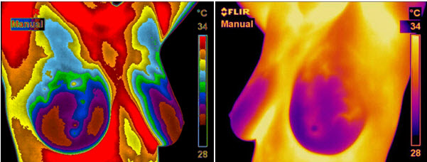
i.Thermography Images of temperature variations of breast. (Source - breastthermography.com)
The method uses software interface for breast cancer screening and detection.The visualization screen displays at least thermal image of one breast of the subject.The pixels with highest temperature value being displayed in a first color and having a lowest temperature value being displayed in a second color. The pixels with temperature values between said lowest and highest temperature values being displayed in gradations of color between said first and second colors.
(US Patent: US20160283658)
Several methods like ultrasound , MRI,mammography and other structural imaging tools rely on finding the physical tumor,DII uses the heat produced by increased blood vessel circulation and metabolic changes associated with a tumor’s genesis and growth for detection of cancer cells.Thus if there exists any minute changes in the normal blood vessel activity,the thermal signs in Infrared imaging would suggest that a pre-cancerous state of the breast or the presence an early tumor that is not yet large enough to be detected by physical examination, mammography, or other types of structural imaging.The breast differences which are caused for women who are on hormone replacement, nursing or have fibrocystic, large, dense, or enhanced breasts etc which would be difficult to read in mammograms but not in DII scans. (breastthermography.com)
PRESSING ISSUES IN DETECTING CANCER:
The current gold standard for breast cancer screening, Mammography, requires high capital cost for equipment and experienced radiographers. It is recommended once every 2 years and only to women above 45 years because it cannot identify tumours effectively for younger women, and uses of X-rays for scanning, which can make women more susceptible to cancer if screened multiple times. In addition, it is also very painful for the subject (about 20 pounds weight is applied on the breasts while screening). Other common method of clinical exam can detect tumors only after they are large enough to be palpable. (niramai.com)
Problems persists with trained radiologists and thermographers looking for the abnormalities in thermal images to make a decision whether tissue is cancerous or is suspicious of being cancerous. Further if found doubtful they may undergo additional tests, such as sonomammography followed by cancer diagnosis through histopathology by fine needle aspiration cytology or tissue biopsy. Both the Thermographers and radiologists are urging for more powerful visualization software interface tools to assist them.(US Patent: US20160283658).Moreover, trained medical practitioners in the field of thermography are not readily available in rural areas, automatic screening tools will help open up this market for software applications for breast cancer screening and detection. Accordingly, the state of art for increasing the software tools which enable subjects as well as medical practitioners to manually or automatically analyze a thermal image of an area of breast tissue for the presence of cancerous tissue if needed.(niramai.com)
ADVANCEMENTS IN DETECTION ACCURACY FOR BREAST CANCER: SOLUTION FROM INDIAN STARTUP
Under the initiative of Make in India, startups are coming with innovation in breast cancer detection as an improvement and development in thermography. An Indian Startup came with a novel and affordable technique to diagnose and detect breast cancer at early stage. NIRAMAI Health Analytix Pvt Ltd have developed new cancer screening software that uses machine intelligence over thermography images to enable a low cost, easy to use, portable solution which requires minimal human supervision. Niramai uses a high-resolution thermal sensing device that scans the chest area like a camera. It then uses cloud-hosted analytics solution for analyzing the thermal images. Its SaaS solution uses big data analytics, artificial intelligence and machine learning for reliable, early and accurate breast cancer screening. (bellaward.com)
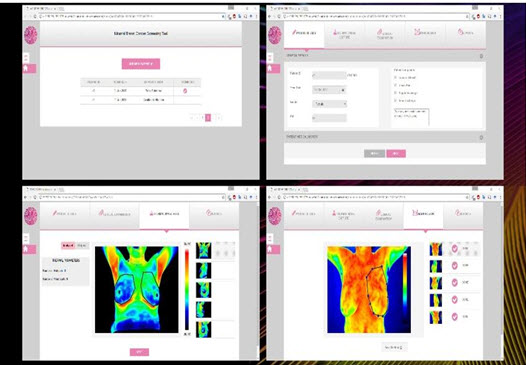
ii. SMILE interface of Niramai showing thermal image uploading platform to determine presence of cancer(Source-Niramai.com)
Their cancer screening tool named SMILE, is an interface for the user to upload the information about patient and thermal images. The information is processed using their patented technology, thermalytix, to analyse the breast health condition. Early results from these tests indicate very high accuracy that is comparable and sometime better that Mammography. This enables many more women to comfortably undergo the test without anyone seeing them or touching them during the test. Over 1700 patients have been screened using NIRAMAI technology with high levels of accuracy. It is effective for decision making. (Niramai.com)
CONCLUSION
The intervention of Niramai ‘s technique will add feathers in effective and accurate finding of Breast Cancer for effective decision making. Primarily both physical examination and Mammography were used to detect suspicious signs of breast cancer .Since the absolute prevention of breast cancer has not become a reality as of yet, efforts must be directed at detecting breast cancer at its earliest stage. Along with Mammography as such, the addition of Digital Infrared Imaging (Breast Thermography) to the frontline of early breast cancer detection brings a great deal of good news for women.The method can detect malignancy at a much earlier stage than traditional diagnostic methods and self-examination, so can therefore improve survival rates. This screening can detect tumors 5 times smaller than what clinical exam can detect, is non-contact, painless and free of any radiation, apart from being low-cost, and universally accessible. With this solution, women of all age groups can undergo frequent screening without any side-effects.
REFERENCES:
1. American Cancer Society – Breast Cancer Guidelines and Statistics, (2009-2010) Available at:(https://www.cancer.org/content/dam/cancer-org/research/cancer-facts-and-statistics/breast-cancer-facts-and-figures/breast-cancer-facts-and-figures-2009-2010.pdf)
2. C. Gros, M.D., M. Gautherie, Ph.D.(1980); “Breast Thermography and Cancer Risk Prediction. Cancer”; 45(1); 51-56.
3. I. Nyirjesy, M.D. et al (1986) ; Clinical Evaluation, Mammography and Thermography in the Diagnosis of Breast Carcinoma. Thermology; 1: 170-173.
4.Indian Startup – Niramai.Available at: https://yourstory.com/2017/07/healthtec-startup-niramai-breast-cancer/
5. J. Keyserlingk, M.D.(1997); "Time to Reassess the Value of Infrared Breast Imaging?" Oncology News Int.; 6(9).
6. M. Gautherie, Ph.D.(1983); "Thermobiological Assessment of Benign and Malignant Breast Diseases". Am. J. Obstet. Gynecol. ;147(8): 861-869
7. Niramai,Breast cancer detection .Available at http://niramai.com
8. Niramai Health Analytix:Available at http://bellaward.com/niramai-health-analytix-wins-aegis-graham-bell-awards/
9.N. Belliveau, M.D., J. Keyserlingk, M.D. et al (1998); "Infrared Imaging of the Breast: Initial Reappraisal Using High-Resolution Digital Technology in 100 Successive Cases of Stage I and II Breast Cancer". Breast Journal ; 4(4)
10.P.Ahlgren, M.D., E. Yu, M.D., J. Keyserlingk, M.D.(1998); "Is it Time to Reassess the Value of Infrared Breast Imaging?" Primary Care & Cancer (NCI);18(2)
11. P. Haehnel, M.D., M. Gautherie, Ph.D. et al (1980); "Long-Term Assessment of Breast Cancer Risk by Thermal Imaging." In: Biomedical Thermology, ; 279-301.
12.P. Gamigami, M.D.(1996); "Atlas of Mammography: New Early Signs" in Breast Cancer. Blackwell Science, United States.
13. Thermography: Breast thermography, available at: http://www.breastthermography.com
14.US Patent: US20160283658 of Niramai Health Analytix Pvt Ltd.
NOW YOU CAN ALSO PUBLISH YOUR ARTICLE ONLINE.
SUBMIT YOUR ARTICLE/PROJECT AT editor-in-chief@pharmatutor.org
Subscribe to Pharmatutor Alerts by Email
FIND OUT MORE ARTICLES AT OUR DATABASE