

{ DOWNLOAD AS PDF }
ABOUT AUTHOR:
*1Patitapabana Parida, 1Bibhukalyan P Nayak, 2Subash Chandra Mishra
1Department of Biotechnology and Medical Engineering,
2Department of Metallurgy & Materials Engineering
National Institute of Technology Rourkela, Odisha, India
*paridap@nitrkl.ac.in
ABSTRACT
Nano structure possesses high surface area that the functional groups can be able to expose their reactive capacity due to high surface energy. So as to consider their size of the amphiphilic nanoparticle dual-surface structure containing body called as janus particle introduced since about twenty years in industry with purpose of drug delivery and diagnostic purpose in the field of paramedical areas. Janus bodies can carry both lipophilic and hydrophilic groups on its surface. Nano-sized morphology could play multiple roles in cancer treatment including diagnosis, biomedical imaging, tissue engineering and drug delivery.
INTRODUCTION
Janus nanoparticle having two surfaces contains both hydrophilic as well as a hydrophobic group. These are unique types of nanoparticles those surfaces have two or more distinct physical properties of chemistry in same molecule[1]. Asymmetric geometry of anisotropic (Janus) nanoparticles considered to either shape or surface composition have fascinated incredible attention due to their optical, magnetic, and surface properties[2]. As these particles contains both hydrophilic and hydrophobic faces are similar to molecular surfactants. The chemical nature of particles can be use as effective and potential stabilizers for a variety of multi-segment systems such as emulsions and foams. Particles may have either an anisotropic or an anisotropic structure depending on distribution of functional groups[3].
Methods of Formulation
There were different techniques used for the fabrication of janus nanoparticles with the help of organic, in-organic, cross-linking phase separation, in the presence of ionic radicals, by metal and salt as catalyst, application of electrostatic force e.t.c by varing in the concentration. Typically designed Janus inorganic hollow spheres were compartmentalized onto both interior and exterior surfaces where inorganic cages posed cross-linked micelles. In brief, organic components could be imbibed inside the cavity while the hydrophilic surface could ensuring dispersal of the hollow spheres in water[4]. Nanosized dual-face Janus particles could formulated by ligand exchange reactions of a Langmuir monolayer of hydrophobic alkanethiolate passivated gold nanoparticles at relatively high surface pressures where with hydrophilic thiol derivatives injected into the water subphase. Interfacial mobility of the particles can resisted the ligand intercalation between adjacent particles. Ligand place-exchange reactions were limited only to the side of the particles facing the water phase, hence leading to the formation of amphiphilic nature of nanoparticles which exhibited hydrophobic characters on one side and hydrophilic on the other[5]. Self assembled block copolymers, and mixtures of ligands that in some cases show competitive adsorption on the surfaces of the nanoparticles is the first deposit where second deposit comprises amphiphile obtained by masking step, where particles could be trap at the interface between two phases, so that a alteration to the constituent surface is made only on one side[6]. Nano sized colloids with dual faced amphiphilic particles with altered chemical composition, acquire energetic interactions depends not only on their separation but also on their orientation[7]. The transfer of positively charged nanoparticles, nanorods and neutral silica nanoparticles into organic solutions could possible to coated and were rely on ligand exchange and this novel method allows the nanoparticles to retain their negative charge[8]. One hydrophilic segment and two incompatible hydrophobic segments were able to made composite in same nano-structured to amphiphilic terpolymers could readily undergo microphase separation where self assembled micelles exposed to aqueous environment. Thus design and generation of discrete multicompartment with hierarchical nanostructures attempted to introduce hydrocarbon and fluorocarbon hydrophobic segments into linear dendritic amphiphilic A-graft-B-block-A-graft-C copolymers[9]. Method like microfluidic and flow-lithography techniques could used to produce Janus particles with good control over size distribution limited to a diameter of approximately 5–100 nm and different shapes[10]. Some forms of janus particle i.e magnetic nanoparticles (MNPs) containing active compounds has been of great concern specially in bio-medical applications[11].
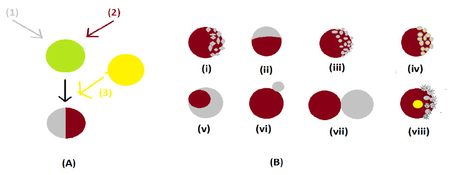
Figure 1. Mismatched compounds (1, 2 with 3) were fixed to form complex nanoparticle (A), various forms of janus particle (B)
Applications in drug delivery
Controlled size and morphology of nanoparticle has great and essential properties for the preparation of many drug delivery devices. The vital properties of nanoparticles could be obtained by using various methods like solvent extraction, solvent diffusion and nano-precipitation in the field of novel drug delivery or, site specific drug delivery[12]. The biphasic nature of amphiphile allows temporally controlled release of multiple curative and diagnostic agents in a specific categorize and with self-determining release rates. Biodegradable Janus particles could be synthesized from a 2.5% w/v solution of PLGA: PCL (50/50 weight ratio) or PLGA: Precirol® (75/25 weight ratio) in dichloromethane (DCM) emulsified into an aqueous solution containing 0.3% w/v PVA and 0.1% w/v sodium dodecyl benzylsulfate (SDBS). Water insoluble drugs were dissolved in the oil phase along with the polymers and lipids, even as water soluble drugs are adsorbed onto the particle surface post-synthesis i.e emulsification and homogenization. Where the higher the energy applied in the emulsification step, the smaller the emulsion droplet[13]. Patchy particles could synthesized be via a simple colloidal route through surfactant free cationic polystyrene nanospheres as core particles where gold patches were composed by in situ diminution of chloroauric acid with ascorbic acid. The nanostructured metal patches could be heterogeneously nucleated on polymer nanospheres is related to the electrostatic interaction between core and metal precursor [14]. The HTSC development of substituted PLA with opposite configurations could provide another versatile way to prepare poly(2-hydroxyalkanoic acid)-based biodegradable materials having a broad variety of physical properties and biodegradability[15]. Two dimensional designs of Janus disk containing of a hydrophobic semicircle and an electro-negatively charged one when placed in a solution, the hydrophobic sides will attract each other while the charged sides will give rise to a repulsive force. The morphology of some particles performed a one dimensional harmonic confinement potential in a channel-like environment whether simulated under molecular dynamics. The interest of system is could serve as a simple model for membrane formation acting as Janus dendrimers a new class of amphiphiles and were revealed to self assemble in bilayer structures mimicking biological membranes [16]. A superparamagnetic Janus nanocomposite of polystyrene/Fe3O4@SiO2 was designed and developed with dual surface containing functional groups for medical diagnosis and treatment. Folic acid and doxorubicin could be arranged stepwise to the surfaces. These particles found to enable simultaneous cell-targeted drug delivery via pH-responsive release mechanism[17]. Poly(ε-caprolactone)-based multicompartment micelles with adjustable Janus-cores could be formed with well-defined terpolymers having A-block-B-graft-C construction framed with biologically compatible polymers i.e methoxy poly(ethylene glycol), poly(ε-caprolactone) and poly(2-(perfluorobutyl)ethyl methacrylate) were synthesized by the stepwise apply of ring opening polymerization and atom transfer radical polymerization. Excellent cytocompatibility could be found in vitro cell viability assays performed of the MCMs both in mouse embryonic fibroblasts and human acute monocytic leukemia cells[23]. Polymeric nanoassemblies had of multiple polymeric building blocks with hierarchal structure and enhanced functionalities would be discussed to create multifunctional nanoparticles by combination of the designing principles which could to some extent tackle challenges widely accessible in nanomedicine such as long blood circulation, efficient cellular uptake, and controllable release of payloads [18]. In the case of dhesion failure and skin reaction due to the occlusive properties of hydrophobic pressure sensitive adhesives, transdermal drug patch based on self-adhesive Janus nanofibrous film was prepared by a multilayered electrospinning which were bilayer structure. The layer was a hydrophobic and adhesive fibrous layer electrospun from polyacrylate and the upper backing layer was a hydrophilic cross-linked poly (vinyl alcohol) nanofibrous film. The structures of the HPSA/c-PVA composite fibrous films were characterized and their application properties, including adherence performance, water vapor permeability, water-penetration, release characteristics, and skin irritation were evaluated [19]. Some Novel classes of jellyfish-shaped amphiphilic dendrimers made of 7 hydrophilic poly ethylene glycol arms and 14 hydrophobic polyester dendrons with β-cyclodextrin as the core molecule were fabricated by a facile method. Seven PEG chains were first conjugated to the C-6 positions of native β-cyclodextrin. Subsequently, dendritic polyester architectures were constructed from the remaining 14 secondary hydroxyl groups at C-2 and C-3 positions of the βCD moiety, resulting in jellyfish-shaped amphiphilic dendrimers of different generations (7PEG-βCD-Gx) with well-defined molecular structures[20].
Applications in diagnostic purpose
Colloidal particles have solid supports of biomolecules for cell separation, as contrast agents in detection tools for in vivo biomedical diagnostic, as support for sample preparation of nucleic acids extraction, and as nanocapsules for active biomolecules in drug delivery systems. Janus colloidal particles had attracted significant attention due to their novel morphologies and diverse potential applications in various domains including materials science, biomedicine and in the field of highly specific biosensors[21]. Polymeric, inorganic, and polymer inorganic, Janus magnetic polymeric nanoparticles had attracted great interest for both fundamental studies and their potential use in applications such as multimodal probes, biosensors, and building blocks in the fabrication of complex nanoscale devices[2]. A variety of fluorescent-magnetic Janus particles (F-MJPs) with complex shapes, anisotropic nature, and diverse functionalities, have been developed by several methods, e.g., microfluidic device and solvent evaporation methods. Although the microfluid device produces a large amount of F-MJPs in a continuous fashion, the size of resultant particles is typically large (1–100 lm)[11]. Anisotropic polymeric colloidal Janus particles possessing simultaneous magnetic and fluorescent properties were successfully prepared via the swelling-diffusion. The in situ emulsionpolymerization method as the swelling-diffusion process, magnetic emulsions (an organic ferro-fluid dispersed in aqueous medium were fabricated and used for seeds of submicron magnetic Janus particles. After swelling the anisotropic particles obtained by 1-pyrene-carboxaldehyde fluorescent dye dissolved in tetrahydrofuran, well-defined fluorescent-magnetic Janus particles were produced. In the in situ emulsion polymerization, styrene monomer mixed with fluorescent dye monomers, i.e., 1-pyrenylmethyl methacrylate or fluorescein dimethacrylate, and an oil-soluble initiator (2,2′-azobis(2-isobutyronitrile) were emulsified in the presence of magnetic seed emulsions [24].Biocompatibility and potential biomedical applications of the Au @ MnO @ SiO2 Janus particles were assayed by a cell viability analysis by coincubating the Au @ MnO @ SiO2 Janus particles with Caki 1 and HeLa cells. Time-resolved fluorescence spectroscopy in combination with CLSM revealed the silica-coated Au @ MnO @ SiO2 Janus particles to be highly two-photon active; no indication for an electronic interaction between the dye molecules incorporated in the silica shell surrounding the MnO domains and the attached Au domains was found; fluorescence quenching was observed when dye molecules were bound directly to the Au domains[22].
CONCLUSION
The article performes the various amphiphilic janus particles those having different characteristic properties. The dual faced particle had role to play as drug delivery, diagnostic and other applications in the field of biomedical and therapeutic consideration. We can use the amphiphilic particles in the field of delivery anticancer agents to specific tissue. Nano sized structure can be used in the field of tissue engineering.
REFERENCES
1. Wikipedia F. Janus particles. 2014.
2. Kaewsaneha C, Tangboriboonrat P, Polpanich D, Eissa M, Elaissari A. Facile method for preparation of anisotropic submicron magnetic Janus particles using miniemulsion. J Colloid Interface Sci. 2013;409:66-71.
3. Ali N, Zhang B, Zhang H, Zaman W, Li W, Zhang Q. Key synthesis of magnetic Janus nanoparticles using a modified facile method. Particuology. 2014:1-7.
4. Li J, Wang Y, Zhang C, et al. Janus polymeric cages. Polymer (Guildf). 2012;53(17):3712-3718.
5. Pradhan S, Xu L, Chen S. Janus Nanoparticles by Interfacial Engineering. Adv Funct Mater. 2007;17(14):2385-2392.
6. Lattuada M, Hatton TA. Synthesis, properties and applications of Janus nanoparticles. Nano Today. 2011;6(3):286-308.
7. Jiang S, Chen Q, Tripathy M, Luijten E, Schweizer KS, Granick S. Janus particle synthesis and assembly. Adv Mater. 2010;22:1060-1071.
8. Allows IA, Snowmen J, Home S, Map S, Us C. Innovative Approach Allows “ Janus Snowmen ” to be Synthesized. 2014:1-2.
9. Wang W, Zhang J, Li C, et al. Facile access to cytocompatible multicompartment micelles with adjustable Janus-cores from A-block-B-graft-C terpolymers prepared by combination of ROP and ATRP. Colloids Surf B Biointerfaces. 2014;115:302-9.
10. Champion J a, Katare YK, Mitragotri S. Particle shape: a new design parameter for micro- and nanoscale drug delivery carriers. J Control Release. 2007;121(1-2):3-9.
11. Kaewsaneha C, Bitar A, Tangboriboonrat P, Polpanich D, Elaissari A. Fluorescent-magnetic Janus particles prepared via seed emulsion polymerization. J Colloid Interface Sci. 2014;424:98-103.
12. Furlan M, Kluge J, Mazzotti M, Lattuada M. Preparation of biocompatible magnetite–PLGA composite nanoparticles using supercritical fluid extraction of emulsions. J Supercrit Fluids. 2010;54(3):348-356.
13. Search Meeting At-A-Glance Browse ... Create an Itinerary AIChE Home Page Google Yelp Facebook Twitter ChEnected. 2014:337461.
14. Bao H, Bihr T, Smith A-S, Klupp Taylor RN. Facile colloidal coating of polystyrene nanospheres with tunable gold dendritic patches. Nanoscale. 2014;6(8):3954-66.
15. Tsuji H, Hayakawa T. Hetero-stereocomplex formation between substituted poly(lactic acid)s with linear and branched side chains, poly(l-2-hydroxybutanoic acid) and poly(d-2-hydroxy-3-methylbutanoic acid). Polymer (Guildf). 2014;55(3):721-726.
16. Ghosh PK, Misko VR, Marchesoni F, Nori F. Self-Propelled Janus Particles in a Ratchet: Numerical Simulations. Phys Rev Lett. 2013;110:268301.
17. Wang F, Pauletti GM, Wang J, et al. Dual surface-functionalized Janus nanocomposites of polystyrene/Fe2O2@SiO2 for simultaneous tumor cell targeting and stimulus-induced drug release. Adv Mater. 2013;25:3485-9.
18. Zhang Z, Ma R, Shi L. Cooperative Macromolecular Self-Assembly toward Polymeric Assemblies with Multiple and Bioactive Functions. Acc Chem Res. 0(0):null.
19. Shi Y, Li Y, Wu J, Wang W, Dong A, Zhang J. A novel transdermal drug delivery system based on self-adhesive Janus nanofibrous film with high breathability and monodirectional water-penetration. J Biomater Sci Polym Ed. 2014;25(7):713-728.
20. Shao S, Si J, Tang J, Sui M, Shen Y. Jellyfish-Shaped Amphiphilic Dendrimers: Synthesis and Formation of Extremely Uniform Aggregates. Macromolecules. 2014;47(3):916-921.
21. Kaewsaneha C, Tangboriboonrat P, Polpanich D, Eissa M, Elaissari A. Preparation of Janus colloidal particles via Pickering emulsion: An overview. Colloids Surfaces A Physicochem Eng Asp. 2013;439:35-42.
22. Schick I, Lorenz S, Gehrig D, et al. Multifunctional Two-Photon Active Silica-Coated Au@MnO Janus Particles for Selective Dual Functionalization and Imaging. J Am Chem Soc. 2014;136:2473-83.
23. Wang, Weiwei, et al. "Facile access to cytocompatible multicompartment micelles with adjustable Janus-cores from A-< i> block</i>-B-< i> graft</i>-C terpolymers prepared by combination of ROP and ATRP." Colloids and Surfaces B: Biointerfaces 115, 2014: 302-309.
24. Gao, Jinhao, et al. "Fluorescent magnetic nanocrystals by sequential addition of reagents in a one-pot reaction: a simple preparation for multifunctional nanostructures." Journal of the American Chemical Society 129.39, 2007: 11928-11935.
REFERENCE ID: PHARMATUTOR-ART-2208
|
PharmaTutor (ISSN: 2347 - 7881) Volume 2, Issue 7 Received On: 14/05/2014; Accepted On: 20/05/2014; Published On: 01/07/2014How to cite this article: P Parida, BP Nayak, SC Mishra; Amphiphilic Janus like Particles for Biomedical Application; PharmaTutor; 2014; 2(7); 92-97 |
NOW YOU CAN ALSO PUBLISH YOUR ARTICLE ONLINE.
SUBMIT YOUR ARTICLE/PROJECT AT articles@pharmatutor.org
Subscribe to Pharmatutor Alerts by Email
FIND OUT MORE ARTICLES AT OUR DATABASE


