{ DOWNLOAD AS PDF }
ABOUT AUTHORS
Devender Sharma*, Dr. Satish Kosalge1, Snehal R. Dixit1
Hi – Tech college of pharmacy, Chandrapur (M.S.)
*sdevender350@gmail.com
ABSTRACT
Aging is one of the phases of individual’s life span, which everyone wants to escape. The process of aging is much more a social phenomena than biological phenomena. Aging brings many changes such as, loss of eyesight, hearing loss, dementia, etc. Aging may give rise to diseased states such as, heart disease, cancer, cerebrovascular disease (relating to blood vessels that supply the brain), pneumonia and flu, and chronic obstructive pulmonary diseases. The key-factor behind aging is the GDF11, i.e., Growth and Differentiation factor-11. GDF11 is also known as Bone Morphogenetic Factor-11 (BMP11). It is termed as key circulating ‘anti-aging’ factor. GDF11 is expressed in wide range of tissues and has been shown to play important roles in development of olfactory system, retina and pancreas. It functions in regulating anterior-posterior patterning of the axial skeleton muscle system. Scientists theorize that aging likely results from a combination of many factors viz., Lifestyle, Diseases and Genes. GDF11 is essential for mammalian development and has been suggested to regulate aging of multiple tissues, whereas myostatin is a well-described negative regulator of postnatal skeletal and cardiac muscle mass and modulates metabolic processes .
[adsense:336x280:8701650588]
Reference Id: PHARMATUTOR-ART-2546
|
PharmaTutor (Print-ISSN: 2394 - 6679; e-ISSN: 2347 - 7881) Volume 5, Issue 12 Received On: 18/08/2017; Accepted On: 18/08/2017; Published On: 01/12/2017 How to cite this article: Sharma D, Kosalge S, Dixit SR;Influence of GDF-11 in Aging Process : An Review; PharmaTutor; 2017; 5(12); 18-29 |
INTRODUCTION
A life course is the period from birth to death, including a sequence of predictable life events such as physical maturation and the succession of age-related roles: child, adolescent, adult, parent, senior etc. Aging is one of the phases of individual’s life span, which everyone wants to avoid. The process of aging is much more a social phenomena than biological phenomena. Some people fear old age and do many things to avoid it, seeking medical and cosmetics remedies for the natural effects of age. Normally, aging brings many changes such as, loss of eyesight, hearing loss, dementia, etc. Aging may give rise to diseased states such as, heart disease, cancer, cerebrovascular disease (relating to blood vessels that supply the brain), pneumonia and flu, and chronic obstructive pulmonary diseases. Numbers of psychological problems associated with aging are dementia, depression, anxiety, paranoia, dread, apprehension, sleep problems, behavioral disorders and most commonly Alzheimer’s disease.[1]
Scientists theorize that aging likely results from a combination of many factors viz., Lifestyle, Diseases and Genes. Lifestyle changes and disease condition are the consequences of aging. The key-factor behind aging is the GDF11, i.e., Growth and Differentiation factor-11. GDF11 is also known as Bone Morphogenetic Factor-11 (BMP11). It is termed as key circulating ‘anti-aging’ factor. [1] It is a member of TGF-β super-family and is derived along with myostatin (GDF-8). GDF11is a protein that, in humans is encoded by the gene GDF11. [3] It acts as a cytokine and its paralog is MSTN gene. [3] It is a myostatin-homologous protein that acts as an inhibitor of nerve tissue growth. [4][17] Growth differentiation factor 11 (GDF11) and myostatin (or GDF8) are closely related members of the transforming growth factor β superfamily and are often perceived to serve similar or overlapping roles.[5][6] Both GDF11 and myostatin are synthesized as precursor molecules where an N-terminal pro-domain is cleaved from a C-terminal signaling or mature domain by a furin protease enzyme.[15] GDF11 is essential for mammalian development and has been suggested to regulate aging of multiple tissues, whereas myostatin is a well-described negative regulator of postnatal skeletal and cardiac muscle mass and modulates metabolic processes.[15] In 2014, GDF11 was described as an anti-aging factor in two publications based on results of parabiosis experiments using mice. [5][6] Many other later studies questioned these findings. [7][8][9][10] The actual relationship between GDF11 and the aging continues to be researched. Both GDF11 and myostatin predominantly use the type II receptors activin receptor kinase II-A and type II receptors activin receptor kinase II-B and the type I receptors activin receptor-like kinase 4 (ALK4) and ALK5 to elicit signal transduction via SMAD2 & SMAD3.[11][15][16][17][18] GDF11 also can signal through an additional type I receptor, ALK7.[20] Signaling by GDF11 and myostatin is regulated by extracellular-binding proteins that are typically thought to function as antagonists. These include follistatin, follistatin-like 3 (FSTL3), decorin, and growth/differentiation factor–associated serum proteins 1 and 2 (GASP1 and GASP2).[15][18] GDF11 is involved in cell growth and differentiation, mesodermal formation and neurogenesis (during embryonic development) as well as in cardiac and skeletal muscle aging (anti-hypertrophic effect and anti-rejuvenating effect). [20][21][22][15] GDF11 is expressed in wide range of tissues and has been shown to play important roles in development of olfactory system, retina and pancreas.[18][26][27][28][29] It functions in regulating anterior-posterior patterning of the axial skeleton muscle system. [33][30]
WHAT IS AGEING?
The aging process happens during an individual’s lifespan. We all are involved in this process and no one can escape it. Many people fear old age and do anything to avoid it. The process of aging is a lifelong process and entails maturation and changes in physical, psychological and social patterning of life. Dr. Ignatz Nascher, a New York Physician gave the term geriatrics, a combination of two Greek words: geron (old man) and iatrikos (medical treatment). He saw the practice of caring for the elderly as separate from the practice of caring for the young, just as pediatrics (caring of children) is different from caring for grown adults.[36] When one is young, aging is associated with growth, maturation, and discovery. In the broader sense, ageing can refer to single cells within an organism which have ceased dividing (cellular senescence) or to the population of a species (population ageing).In humans, aging represents the accumulation of changes, encompassing physical, psychological, and social changes.[57] Ageing is among the greatest known risk factors for most human diseases:[48] roughly 150,000 people who die each day across the globe, about two thirds die from age-related causes.
Scientists theorize that aging likely results from a combination of many factors. Genes, lifestyle, and diseases can all affect the rate of aging. Studies indicate that people age at different rates and in different ways.[56]

Fig.No.1: A 75 year old woman
Effects of aging
A number of characteristic ageing symptoms are experienced by a majority or by a significant proportion of humans during their lifetimes. Normal aging brings about following changes:
• Eyesight- loss of vision or blurred vision and decreased ability to identify objects.[59][60] More than half, may undergo cataract surgery. [65]
• Hearing- loss of hearing acuity and decreased ability to distinguish sounds when there is background noise.[64]
• Taste- decreased taste buds and saliva.
• Touch and Smell- decreased ability to smell and sensitivity to touch.
• Arteries- hardening of the arteries, leading to Arteriosclerosis. [68]
• Brain- loses some of the structures that connect nerve cells, thus, diminishing cell functions. High risk of dementia, anxiety, apprehension and behavioral disorders etc.[71][72]
• Bones- lose minerals leading to osteoarthritis. [63]
• Bladder- increased frequency in urination.
• Heart- pumping rate and body’s ability to extract blood diminishes with age. Also higher risk of stroke and heart attacks. [68][69][70]
• Kidneys- shrink and become less efficient.
• Lungs- lung tissue begins to lose its elasticity and bronchial muscle shrink, thus, decreasing breathing capacity.
• Muscles- muscle mass decline. [66][67]
• Skin- nails grow more slowly. Skin is more dry and wrinkled.
• Sexual health- women go through menopause, vaginal lubrication decreases. In men, sperm production decreases and the prostate enlarges. Hormone level decreases.[62][73]
• Hairs- hair turns grey with age. [61]

Fig. No. 2: An old woman with wrinkled face and grey colored hairs
NOW YOU CAN ALSO PUBLISH YOUR ARTICLE ONLINE.
SUBMIT YOUR ARTICLE/PROJECT AT editor-in-chief@pharmatutor.org
Subscribe to Pharmatutor Alerts by Email
FIND OUT MORE ARTICLES AT OUR DATABASE
The process of aging
Aging is not only the physiological process but also a social and biological. The fact that age-related roles and identities vary according to social determinations actually means that the aging is much more a social than a biological phenomena. Riley, scientists (1978) noted that aging is a lifelong process that involves change in physical, psychological, and social levels. Aging can be visible, public experience. Many people recognize the signs of the aging, and believes that being older means being in a physical decline. Each person experiences age-related changes based on many factors.
Biological Basis of aging
Biological factors such as molecular and cellular changes are called primary aging, while aging that occurs due to controllable factors such as lack of exercise and poor diet is called secondary aging. Factors that are proposed to influence biological aging [74] fall into two main categories, programmed and damage-related. Programmed factors may include changes in gene expression that affect the systems responsible for maintenance, repair and defence responses. Damage-related factors include internal and environmental assaults to living organisms that induce cumulative damage at various levels.[75] There are three main metabolic pathways which can influence the rate of ageing:
• The FOXO3/Sirtuin pathway, probably responsive to caloric restriction
• The Growth hormone/Insulin-like growth factor 1 signaling pathway
• The activity levels of the electron transport chain in mitochondria.[76]
It is likely that most of these pathways affect ageing separately, because targeting them simultaneously leads to additive increases in lifespan.
DNA damage theory of ageing
DNA damage is thought to be the common basis of both cancer and ageing, and it has been argued that intrinsic causes of DNA damage are the most important drivers of ageing.[77][78][79] Genetic damage (aberrant structural alterations of the DNA), mutations (changes in the DNA sequence), and epimutations (methylation of gene promoter regions or alterations of the DNA scaffolding which regulate gene expression), can cause abnormal gene expression. DNA damage causes the cells to stop dividing or induces apoptosis, often affecting stem cell pools and hence hindering regeneration. Genetic damage (particularly gene loss) is the most probable cause of aging. [80]
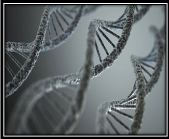
Fig. no.3: The double stranded DNA
How Genes Impact Lifespan
It doesn't take an elaborate study to determine that our genes play at least some role in longevity. People, whose parents and ancestors have lived longer, tend to live longer and vice versa. At the same time, we know that genetics alone are not the sole cause of aging. Studies looking at identical twins reveal that there is clearly something else going on; identical twins who have identical genes do not always live an identical number of years.
Some genes are beneficial and enhance longevity. For example, the gene that helps a person metabolize cholesterol would reduce a person's risk of heart disease.
Some gene mutations are inherited, and may shorten lifespan. However, mutations also can happen after birth, since exposure to toxins, free radicals and radiation can cause gene changes. (Gene mutations acquired after birth are referred to as acquired or somatic gene mutations.) Most mutations are not bad for you, and some can even be beneficial. That's because genetic mutations create genetic diversity, which keeps populations healthy. Other mutations, called silent mutations, have no effect on the body at all.
Some genes, when mutated are harmful, like those that increase the risk of cancer. Many people are familiar with the BRCA1 and BRCA2 mutations which predispose to breast cancer. These genes are referred to as tumor suppressor genes which code for proteins that control the repair of damaged DNA (or the elimination of the cell with damaged DNA if repair is not possible.)
Various disease and conditions related to heritable gene mutations can directly impact lifespan. These include cystic fibrosis, sickle cell anemia, Tay-Sachs disease and Huntington's disease, to name a few.[80][88]
Key Concepts in the Genetic Theory of Aging
The key concepts in genetics and aging include several important concepts and ideas ranging from telomere shortening to theories about the role of stem cells in aging.
Telomeres - At the end of each of our chromosomes lies a piece of "junk" DNA called telomeres. Telomeres do not code for any proteins but appear to have a protective function, keeping the ends of DNA from attaching to other pieces of DNA or forming a circle. Each time a cell divides a little more of a telomere is snipped off. Eventually, there is none of this junk DNA left, and further snipping can damage the chromosomes and genes so that the cell dies.
In general, the average cell is able to divide 50 times before the telomere is used up . Cancer cells have figured out a way to not remove, and sometimes even add to, a section of the telomere. In addition, some cells such as white blood cells do not undergo this process of telomere shortening. It appears that while genes in all of our cells have the code word for the enzyme telomerase which inhibits telomere shortening and possibly even results in lengthening, the gene is only "turned on" or "expressed" as geneticists say, in cells such as white blood cells and cancer cells. Scientists have theorized that if this telomerase could somehow be turned on in other cells (but not so much that their growth would go haywire as in cancer cells) our age limit could be expanded. [80][88]
Studies have found that some chronic conditions such as high blood pressure are associated with less telomerase activity whereas a healthy diet and exercise are linked with longer telomeres. Being overweight is also associated with shorter telomeres. [88]
Longevity genes - Longevity genes are specific genes which are associated with living longer. Two genes that are directly associated with longevity are SIRT1 (sirtruin 1) and SIRT2. Scientists looking at a group of over 800 people age 100 or older, found three significant differences in genes associated with aging.[88]
Cell senescence - Cell senescence refers to the process by which cells decay over time. This can be related to shortening of the telomeres, or the process of apoptosis (or cell suicide) in which old or damaged cells are removed. [80][88]
Stem cells - Pluripotent stem cells are immature cells which have the potential to become any type of cell in the body. It is theorized that aging may be related to either the depletion of stem cells or the loss of the ability of stem cells to differentiate or mature into different kinds of cells. It's important to note that this theory refers to adult stem cells, not embryonic stem cells. Unlike embryonic stem cells, adult stem cells cannot mature into any type of cell but rather only a certain number of cell types. Most cells in our bodies are differentiated, or fully mature, and stem cells are only a small number of the cells present in the body.
An example of a tissue type in which regeneration is possible by this method is the liver. This is in contrast to brain tissue which usually lacks this regenerative potential. There is now evidence that stem cells themselves may be affected in the aging process, but these theories are similar to the chicken-and-the-egg issue. It's not certain of aging occurs due to changes in stem cells, or, if instead, changes in stem cells are due to the process of aging. [88]
Epigenetics - Epigenetics refers to the expression of genes. In other words, a gene may be present, but can either be turned on or turned off. We know that there are some genes in the body that are turned on for only a certain period of time. The field of epigenetic is also helping scientists understand how environmental factors may work within the constraints of genetics to either protect or predispose to disease. [88]
Three Primary Genetic Theories of Aging
As noted above, there is a significant amount of evidence that looks at the importance of genes in expected survival. When looking at genetic theories, these are broken down into three primary schools of thought.
1.The first theory claims that aging is related to mutations which are related to long term survival, and that aging is related to the accumulation of genetic mutations which are not repaired. [80][88]
2. Another theory is that aging is related to the late effects of certain genes, and is referred to as pleiotropic antagonism.[88]
3. Yet another theory, suggested based on survival in opossums, is that an environment which poses few hazards to interfere with life expectancy would result in an increase in members who have mutations that slow down the aging process. [88]
NOW YOU CAN ALSO PUBLISH YOUR ARTICLE ONLINE.
SUBMIT YOUR ARTICLE/PROJECT AT editor-in-chief@pharmatutor.org
Subscribe to Pharmatutor Alerts by Email
FIND OUT MORE ARTICLES AT OUR DATABASE
GDF11 (Growth and Differentiation Factor 11)
NAMES:
• Growth Differentiation Factor 11
• Bone Morphogenetic Protein 11
• BMP-11
• GDF-11
• BMP11
Growth and differentiation factor 11 (GDF11) also known as bone morphogenetic protein 11 (BMP11), is a protein that in humans is encoded by the GDF11 gene.[3] It is the member of TGF-β superfamily and is related to GDF8 (Myostatin). [16] It acts as cytokine. GDF11 and GDF8 shares 89% protein sequence homology. [2] GDF11 is believed to play roles in inducing mesoderm in early development and in anterior-posterior patterning of the axial skeleton.[53] Interestingly, GDF11 was recently shown to act as a negative feedback inhibitory signal in neurogenesis of the olfactory epithelium, in a fashion reminiscent of the inhibition of muscle mass by myostatin. Thus, myostatin and GDF11 share structural and functional features. [17][18][53][81] This gene encodes a secreted ligand of the TGF-beta (transforming growth factor-beta) superfamily of proteins. Ligands of this family bind various TGF-beta receptors leading to recruitment and activation of SMAD family transcription factors that regulate gene expression. [1][2][42] The encoded preproprotein is proteolytically processed to generate each subunit of the disulfide-linked homodimer. [16][20] GDF11 also plays an important role in a variety of biological processes including embryonic development, skeleton and muscle formation. Recently, increasing studies showed that GDF11 was closely related to rejuvenation. [1][18] GDF11 was expressed in primitive streak, tail bud region, limbs, mandibular and bronchial arches, dorsal neural tubes, odontoblasts, nasal epithelium and particular regions of the brain, in mice. [16][18][20][21][30][53] Several MSTN and GDF11 binding proteins have been identified, including GDF-associated serum protein-1 (GASP-1) and GASP-2, which are capable of inhibiting the activities of MSTN and GDF11, respectively.[18[23][33] GDF11 can bind type 1 TGF-beta superfamily receptors ACVR1 (ALK4), TGFBR1 (ALK5) and ACVR1C (ALK7), but the actions are mediated more via ALK4 and ALK5 for signal transduction. [11][17[18] GDF11 also inhibits myoblast differentiation via down-regulating muscle-specific genes required for myogenic differentiation. [17]
Genomic Location for GDF11 Gene [53][83]
• Chromosome: 12
• Start: 55,743,278 bp from pter
• End: 55,757,466 bp from pter
• Size: 14,189 bases
GDF11 is activated by the proprotein convertase PCSK5. It was found that teratogenic doses of all-trans retinoic acid (ATRA), when administered to pregnant mice via gavage at embryonic day 9 (E9), inhibited Pcsk5 and Gdf11 expression in the hindgut at E12 and E18. [84]
GDF11 has been proved to be a key circulating ‘anti-aging’ factor. [1][5][6] It was found to circulate in the blood, and declining levels of circulating GDF11 have been reported in the etiology of age-related cardiac hypertrophy. [31][32][43][50] In one of the study,GDF11 has been shown to suppress neurogenesis through a pathway similar to that of myostatin involving stopping the progenitor cell-cycle during G-phase. [4] While the systemic administration of recombinant GDF11 protein restores genomic integrity and health of muscle stem cells, neurovasculature and enhaces neurogenesis. [16][44][45]
Effects of GDF11 on growth and differentiation
GDF11 belongs to TGF-beta superfamily that controls anterior-posterior patterning by regulating the expression of HOX genes. It determines HOX gene expression domains and rostrocaudal identity in the caudal spinal cord.[11]The members of TGF-β superfamily (especially GDF11 and GDF8) are involved in the regulation of cell growth and differentiation in embryonic tissues as well as in adult tissues. GDF11 is specifically involved in mesodermal formation and neurogenesis during embryonic development. [20] GDF11 can bind type1 TGF-β superfamily receptors ACVR1B (ALK4), TGFBR1 (ALK5) and ACVR1C (ALK7), but it mostly uses ALK4 and ALK5 for the signal transduction. [11]
GDF11 is closely related to myostatin, a negative regulator of muscle growth. [12][13] Both myostatin and GDF11 are involved in the regulation of cardiomyocyte proliferation. GDF11 is also a negative regulator of neurogenesis, [3] the production of islet progenitor cells,[82] the regulation of kidney organogenesis,[85] pancreatic development, the rostro-caudal patterning in the development of spinal cords,[11] and is a negative regulator of chondrogenesis.[86]
Effects of GDF11 on cardiac and skeletal muscle aging
GDF11 has been identified as a blood circulating factor that has the ability to reverse age-related cardiac hypertrophy in mice. GDF11 gene expression and protein abundance decreases with age, and it shows differential abundance between young and old mice in parabiosis procedures, causing youthful regeneration of cardiomyocytes, a reduction in the Brain natriuretic peptide (BNP) and in the Atrial natriuretic peptide (ANP). GDF11 also causes an increase in expression of SERCA-2, an enzyme necessary for relaxation during diastolic functions.[46] GDF11 activates the TGF-β pathway in cardiomyocytes derived from pluripotent hematopoietic stem cells and suppresses the phosphorylation of Forkhead (FOX proteins) transcription factors. These effects suggest an "anti-hypertrophic effect", aiding in the reversal process of age-related hypertrophy, on the cardiomyocytes. [46]
In 2014, peripheral supplementation of GDF11 protein (in mice) was shown to ameliorate the age-related dysfunction of skeletal muscle by rescuing the function of aged muscle stem cells, claiming that GDF11 may be an anti-aging rejuvenation factor.[5][52] These previous findings have been disputed since another publication has demonstrated the contrary, concluding that GDF11 increases with age and has deleterious effects on skeletal muscle regeneration, being a pro-aging factor, with very high levels in some aged individuals. [7] In 2015, it was claimed that GDF11 reverse age-related cardiac hypertrophy. [87]
Regulation of GDF-11 & GDF-8 Activity by GASP-1 &GASP-2
GDF-11 and GDF-8 are closely related TGF-β superfamily members, that shares 90% amino acid sequence identity within the mature C- terminal region. [24][25] GDF-8 is expressed in skeletal muscles and act as negative regulator of muscle growth. [24] While, GDF-11 is expressed in a wide range of tissues and has been shown to play an important role in olfactory system [26], retina [27], and pancreas [28] [29] as well as in anterior-posterior patterning of the axial skeleton system. [30]
The regulation of GDF-8 and GDF-11 seems to be complex, since multiple proteins are available that are capable of binding these ligands and inhibits their activities. [31][33] The two main binding proteins responsible for regulation of GDF-8 &GDF-11 are GASP-1 and GASP-2. GASP-1 is also known as WFIKKNRP or WFIKKN2, and it has been identified as MSTN-associated protein. [34] GASP-2 is also known as WFIKKN or WFIKKN1 and has been identified as GDF-11 associated protein.GASP-1 is expressed in skeletal muscles, which is predominant site of MSTN expression, while GASP-2 is expressed in retina, otocyst, neural tube and in the posterior region of brain. [18] Both GASP-1 and GASP-2 shares 54% amino acid sequence similarity. [35][36] It was observed that GASP-1 and GASP-2 are capable of blocking the activities of GDF-8 and GDF-11, respectively. [34][37][38] GASP-1 and GASP-2 mediates their action by blocking the initial binding of these ligands (GDF-8 & GDF11) to its receptors. [18]
It was noted that GASP-1 & GASP-2 are produced as fusion proteins in either COS1 cell or in Dorsophilia S2 cells, and are capable of binding MSTN and GDF11, inhibiting their activities, in vitro. [34][37] Is was observed that mice that lacks GASP-1 and/or GASP-2 exhibits muscle and skeletal phenotypes consistent with overactivity of myostatin and/or GDF-11. [18]
The effect of GASP-1 and GASP-2 on binding of GDF-11 to its high affinity receptors, activin type-2B receptors (ACVR2B), was studied using the purified soluble form of the receptor, ACVR2B/Fc (possessing a Fc domain. It was found that GDF-11 bound readily to ACVR2B/Fc in the absence of either GASP-1 or GASP-2. With the increasing concentration of GASP-1 and GASP-2, the amount of GDF-11 binding to ACVR2B/Fc was decreased in a concentration-dependent manner. Hence, it was concluded that GASP-1 and GASP-2 are capable of blocking the first step in signaling pathway by binding the ligand and inhibiting its ability to engage the type-2 receptor. [18]
Evidences for GDF-11 as an anti-aging factor
HSCI (Harvard Stem Cell Institute) research: [19]
The researchers of HSCI had shown that GDF11 can make failing hearts in aging mice appear more like those of healthy young mice, and also improves the brain and skeletal muscle functions in aging mice. They observed that injecting GDF-11 protein in aged mice (70-years), improved the exercise capability of mice as that and also improved the functions of olfactory regions of the brain of the older mice. This was proved in two ways:
a. Parabiosis experiment, in which two mice are surgically joined and the blood of younger mice circulates through the older mice;
b. By injecting the older mice with GDF-11.
When exposed to the blood of young mice, the enlarged, weakened hearts of the older mice returned to more youthful size, and their functions improved.
Helen Thomson Report: [51]
- This experiment proved that a transfusion of young mouse blood can improve cognition and the health of several organs in older mice, and even make them look younger.
- Heterochronic parabiosis experiment was performed to prove this, in which the circulatory systems of old and young mice were stitched together. It was observed that the cartilage of old mice soon appeared younger.
- Amy Wager, in 2012 proved that young blood can reverse heart decline in old mice. It was proved by pairing the healthy young mice with old mice that had a cardiac hypertrophy (a condition which swells the size of heart)- and thus, connecting their circulatory systems. After 4 weeks, the old mouse’s heart shrunk to the same size as that of young mice; the young mice left unaffected by the old blood.
- The effect of GDF11 to reduce the elevated blood pressure in old mice was proved by injecting the GDF11 protein to the old mice for 30 days.
- The daily injection of GDF11 also increases the number of blood vessels and the number of stem cells in the brain, increasing their functions.
- The physical endurance and cognitive functions also was observed to be improved in old mice after injecting the blood plasma from young mice (possessing high levels of GDF11).
- The old mice were also able to repair injured muscles as well as young mice.
NOW YOU CAN ALSO PUBLISH YOUR ARTICLE ONLINE.
SUBMIT YOUR ARTICLE/PROJECT AT editor-in-chief@pharmatutor.org
Subscribe to Pharmatutor Alerts by Email
FIND OUT MORE ARTICLES AT OUR DATABASE
SUMMARY AND CONCLUSION:
Aging is one of the phases of individual’s life span, which everyone wants to avoid. In humans, ageing represents the accumulation of changes in a human being over time encompassing physical, psychological, and social change. Ageing is among the greatest known risk factors for most human diseases. The causes of ageing are uncertain; current theories are assigned to the damage concept, whereby the accumulation of damage (such as DNA oxidation) may cause biological systems to fail, or to the programmed ageing concept, whereby internal processes (such as DNA methylation) may cause ageing. Programmed ageing should not be confused with programmed cell death (apoptosis). Human beings and members of other species, especially animals, necessarily experience ageing and mortality.
Scientists theorize that aging likely results from a combination of many factors viz., Lifestyle, Diseases and Genes. Lifestyle changes and disease condition are the consequences of aging. The key-factor behind aging is the GDF11, i.e., Growth and Differentiation factor-11. GDF11 is also known as Bone Morphogenetic Factor-11 (BMP11). It is termed as key circulating ‘anti-aging’ factor. It is a member of TGF-β super-family and is derived along with myostatin (GDF-8). GDF11is a protein that, in humans is encoded by the gene GDF11. It acts as a cytokine and its paralog is MSTN gene. It is a myostatin-homologous protein that acts as an inhibitor of nerve tissue growth. Growth differentiation factor 11 (GDF11) and myostatin (or GDF8) are closely related members of the transforming growth factor β superfamily and are often perceived to serve similar or overlapping roles. Both GDF11 and myostatin are synthesized as precursor molecules where an N-terminal pro-domain is cleaved from a C-terminal signaling or mature domain by a furin protease enzyme.
GDF11 is essential for mammalian development and has been suggested to regulate aging of multiple tissues, whereas myostatin is a well-described negative regulator of postnatal skeletal and cardiac muscle mass and modulates metabolic processes .In 2014, GDF11 was described as an anti-aging factor in two publications based on results of parabiosis experiments using mice. Many other later studies questioned these findings. The actual relationship between GDF11 and the aging continues to be researched. Both GDF11 and myostatin predominantly use the type II receptors activin receptor kinase II-A and type II receptors activin receptor kinase II-B and the type I receptors activin receptor-like kinase 4 (ALK4) and ALK5 to elicit signal transduction via SMAD2 & SMAD3 .GDF11 also can signal through an additional type I receptor, ALK7 .Signaling by GDF11 and myostatin is regulated by extracellular-binding proteins that are typically thought to function as antagonists. These include follistatin, follistatin-like 3 (FSTL3), decorin, and growth/differentiation factor–associated serum proteins 1 and 2 (GASP1 and GASP2) .GDF11 is involved in cell growth and differentiation, mesodermal formation and neurogenesis (during embryonic development) as well as in cardiac and skeletal muscle aging (anti-hypertrophic effect and anti-rejuvenating effect). GDF11 is expressed in wide range of tissues and has been shown to play important roles in development of olfactory system, retina and pancreas. It functions in regulating anterior-posterior patterning of the axial skeleton muscle system.
The researchers of HSCI had shown that GDF11 can make failing hearts in aging mice appear more like those of healthy young mice, and also improves the brain and skeletal muscle functions in aging mice (result of Parabiosis experiment). When exposed to the blood of young mice, the enlarged, weakened hearts of the older mice returned to more youthful size, and their functions improved. The young blood can reverse heart decline in old mice. It was proved by pairing the healthy young mice with old mice that had a cardiac hypertrophy (a condition which swells the size of heart) - and thus, connecting their circulatory systems. After 4 weeks, the old mouse’s heart shrunk to the same size as that of young mice; the young mice left unaffected by the old blood. GDF11 also reduces the elevated blood pressure in old mice. The daily injection of GDF11 also increases the number of blood vessels and the number of stem cells in the brain, increasing their functions. The physical endurance and cognitive functions also was observed to be improved in old mice after injecting the blood plasma from young mice (possessing high levels of GDF11). The old mice were also able to repair injured muscles as well as young mice.
Thus it can be concluded that, GDF11 levels decline with increasing age and GDF11 is a key circulating ‘anti-aging’ factor.
REFERENCES
1. Aaron C. Hinken, Janine M. Powers, Guizhen Luo, Jason A, Holt, Andrew N Billin and Alan J. Russel, “Lack of evidence for GDF11 as a rejuvenator of aged skeletal muscle satellite cells”, Aging Cell; 2016 (15), pg. no.: 582-584.
2. Tommaso Poggioli, Ana Vujic, Peiguo Yang, Claudio Macias; “Circulating Growth Differentiation Factor 11/8 Levels Decline with Age”, Circulatory Research; October2015; CIRCRESAHA.115.3075521.
3. Ge G, Hopkins DR, Ho WB, Greenspan DS (Jul 2005). "GDF11 forms a bone morphogenetic protein 1-activated latent complex that can modulate nerve growth factor-induced differentiation of PC12 cells". Molecular and Cellular Biology. 25 (14): 5846–58.Doi: 10.1128/MCB.25.14.5846-5858.2005. PMC 1168807- PMID 15988002.
3. "Recombinant-Human GDF11".
4. Sinha M, Jang YC, Oh J, Khong D, Wu EY, Manohar R, Miller C, Regalado SG, Loffredo FS, Pancoast JR, Hirshman MF, Lebowitz J, Shadrach JL, Cerletti M, Kim MJ, Serwold T, Goodyear LJ, Rosner B, Lee RT, Wagers AJ (May 2014). "Restoring systemic GDF11 levels reverses age-related dysfunction in mouse skeletal muscle". Science. 344(6184): 649–52. doi:10.1126/science.1251152. PMID 24797481.
5. Katsimpardi L, Litterman NK, Schein PA, Miller CM, Loffredo FS, Wojtkiewicz GR, Chen JW, Lee RT, Wagers AJ, Rubin LL (May 2014). "Vascular and neurogenic rejuvenation of the aging mouse brain by young systemic factors". Science. 344 (6184): 630–4. doi:10.1126/science.1251141. PMID 24797482.
6. Egerman MA, Cadena SM, Gilbert JA, Meyer A, Nelson HN, Swalley SE, Mallozzi C, Jacobi C, Jennings LL, Clay I, Laurent G, Ma S, Brachat S, Lach-Trifilieff E, Shavlakadze T, Trendelenburg AU, Brack AS, Glass DJ (Jul 2015). "GDF11 Increases with Age and Inhibits Skeletal Muscle Regeneration". Cell Metabolism. 22 (1): 164–74.doi:10.1016/j.cmet.2015.05.010. PMID 26001423.
7. Age-reversal effects of 'young blood' molecule GDF-11 called into question, retrieved20 May 2015
8. 'Young blood' anti-ageing mechanism called into question, retrieved 20 May 2015
9. Smith SC, Zhang X, Zhang X, Gross P, Starosta T, Mohsin S, Franti M, Gupta P, Hayes D, Myzithras M, Kahn J, Tanner J, Weldon SM, Khalil A, Guo X, Sabri A, Chen X, MacDonnell S, Houser SR (Nov 2015). "GDF11 Does Not Rescue Aging-Related Pathological Hypertrophy". Circulation Research. 117 (11): 926–32.doi:10.1161/CIRCRESAHA.115.307527. PMID 26383970.
10. Andersson O, Reissmann E, Ibáñez CF (Aug 2006). "Growth differentiation factor 11 signals through the transforming growth factor-beta receptor ALK5 to regionalize the anterior-posterior axis". EMBO Reports. 7 (8): 831–7. doi:10.1038/sj.embor.7400752.PMC 1525155. PMID 16845371.
11. McPherron AC, Lee SJ (Nov 1997). "Double muscling in cattle due to mutations in the myostatin gene". Proceedings of the National Academy of Sciences of the United States of America. 94 (23): 12457–61. doi:10.1073/pnas.94.23.12457. PMC 24998 .PMID 9356471.
12. Lee SJ, McPherron AC (Oct 1999). "Myostatin and the control of skeletal muscle mass".Current Opinion in Genetics & Development. 9 (5): 604–7. doi:10.1016/S0959-437X(99)00004-0. PMID 10508689.
13. V. Kh. Khavinsona, b, d, B. I. Kuznikc, S. I. Tarnovskayab, and N. S. Linkovaa, “GDF11 Protein as a Geroprotector” ; ISSN 2079-0864; Biology Bulletin Reviews;2016; vol. 6(2); 141-148.
14. 15. Ryan G. Walker, Tommaso Poggioli, Lida Katsimpardi; December 2016, “Biochemistry and B iology of GDF11 and Myostatin”, Circulation Research,Pg. no.: 1125-1142
15. Recombinanat Human/Mouse/Rat; GDF11/ BMP11; R & D Systems- a biotechne brand; Catalog no. 1958-GD; October 2015.
16. Ferenc Jeanplong, Shelley J. Falconer; “GDF-11 is developmentally regulated in skeletal muscle and inhibits myoblast differentiation”, OJMIP; 2012 (20); Pg. no.: 127-128.
17. Yun- Sil Lee and Se-Jin Lee; “ Regulation of GDF11 and Myostatin activity by GASP1 and GASP2”, Dept. of Molecular Biology and Genetics, Johns Hopkins University School of Medicine, Baltimore, MD 21205; PNAS; 2013; Page No. 3713-3722.
18. Medical Press- “Functioning of aged Brains and Muscles in mice made younger”, Harvard University; May 2014.
19. Gamer, L.W. et al. (1999) Dev. Biol. 208: 222
20. Nakashima, M. et al. (1999) Mech. Dev. 80:185.
21. Gad, J.M. and P.P.L. Tam (1999) Curr. Biol. 9:R783.
22. McPherron, A.C. et al. (1999) Nat. Genet. 22:260.
23. McPherron AC, Lawler AM, Lee SJ (1997) Regulation of skeletal muscle mass in mice by a new TGF-beta superfamily member. Nature 387(6628):83–90.
24. NakashimaM,ToyonoT,AkamineA,JoynerA(1999)Expressionofgrowth/differentiation factor 11, a new member of the BMP/TGFbeta superfamily during mouse embryogenesis. Mech Dev 80(2):185–189.
25. Wu HH, et al. (2003) Autoregulation of neurogenesis by GDF11. Neuron 37(2): 197–207.
26. Kim J, et al. (2005) GDF11 controls the timing of progenitor cell competence in developing retina. Science 308(5730):1927–1930.
27. Harmon EB, et al. (2004) GDF11 modulates NGN3+ islet progenitor cell number and promotes beta-cell differentiation in pancreas development. Development 131(24): 6163–6174.
28. Dichmann DS, Yassin H, Serup P (2006) Analysis of pancreatic endocrine development in GDF11-deficient mice. Dev Dyn 235(11):3016–3025.
29. McPherron AC, Lawler AM, Lee SJ (1999) Regulation of anterior/posterior patterning of the axial skeleton by growth/differentiation factor 11. Nat Genet 22(3):260–264.
30. Souza TA, et al. (2008) Proteomic identification and functional validation of activins and bone morphogenetic protein 11 as candidate novel muscle mass regulators. Mol Endocrinol 22(12):2689–2702.
31. Loffredo FS, et al. (2013) Growth differentiation factor 11 is a circulating factor that reverses age-related cardiac hypertrophy. Cell 153(4):828–839.
32. Lee SJ (2004) Regulation of muscle mass by myostatin. Annu Rev Cell Dev Biol 20: 61–86.
33. Hill JJ, Qiu Y, Hewick RM, Wolfman NM (2003) Regulation of myostatin in vivo by growth and differentiation factor-associated serum protein-1: A novel protein with protease inhibitor and follistatin domains. Mol Endocrinol 17(6):1144–1154.
34. Trexler M, Bányai L, Patthy L (2002) Distinct expression pattern of two related human proteins containing multiple types of protease-inhibitory modules. Biol Chem 383(1): 223–228. 36. Trexler M, Bányai L, Patthy L (2001) A human protein containing multiple types of protease-inhibitory modules. Proc Natl Acad Sci USA 98(7):3705–3709.
35. Kondás K, Szláma G, Trexler M, Patthy L (2008) Both WFIKKN1 and WFIKKN2 have high affinity for growth and differentiation factors 8 and 11. J Biol Chem 283(35): 23677–23684.
36. Szláma G, Kondás K, Trexler M, Patthy L (2010) WFIKKN1 and WFIKKN2 bind growth factors TGFβ1, BMP2 and BMP4 but do not inhibit their signalling activity. FEBS J 277(24):5040–5050.
37. Haidet AM, et al. (2008) Long-term enhancement of skeletal muscle mass and strength by single gene administration of myostatin inhibitors. Proc Natl Acad Sci USA 105(11):4318–4322.
38. Monestier O, et al. (2012) Ubiquitous Gasp1 overexpression in mice leads mainly to a hypermuscular phenotype. BMC Genomics 13(1):541.
39. Esquela, A.F. and S.J. Lee (2003) Dev. Biol. 257:356.
40. Oh, S.P. et al. (2002) Genes & Dev. 16:274.
41. Loffredo, F.S. et. al. (2013) Cell. 153.828.
42. Katsimpardi, L. et. al. (2014) Science (ahead of print).
43. Sinha, M. et. al. (2014) Science (ahead of print).
44. Loffredo FS, Steinhauser ML, Jay SM, Gannon J, Pancoast JR, Yalamanchi P, Sinha M, Dall'Osso C, Khong D, Shadrach JL, Miller CM, Singer BS, Stewart A, Psychogios N, Gerszten RE, Hartigan AJ, Kim MJ, Serwold T, Wagers AJ, Lee RT (May 2013). "Growth differentiation factor 11 is a circulating factor that reverses age-related cardiac hypertrophy". Cell. 153 (4): 828–39. doi:10.1016/j.cell.2013.04.015. PMID 23663781.
45. Lee SJ, McPherron AC. Regulation of myostatin activity and muscle growth. Proc Natl Acad Sci U S A. 2001;98:9306–9311. doi: 10.1073/ pnas.151270098.
46. Andersson O, Reissmann E, Jörnvall H, Ibáñez CF. Synergistic interac- tion between Gdf1 and Nodal during anterior axis development. Dev Biol. 2006;293:370–381. doi: 10.1016/j.ydbio.2006.02.002.
47. Rebbapragada A, Benchabane H, Wrana JL, Celeste AJ, Attisano L. Myostatin signals through a transforming growth factor beta-like signal- ing pathway to block adipogenesis. Mol Cell Biol. 2003;23:7230–7242.
48. Tommaso Poggioli, Ana Vujic, Peiguo Yang; “Circulating Growth and Differentiation 11/8 levels Decline with age”, Circulation Research: Jan. 2017; Pg. no. 1-14.
49. Helen Thomson, “Young Blood Turns Back Time”, New Scientists, 23 August 2014; Pg. no. 8-9.
50. Marc A. Egerman, Samael M. Cadena, Jason A. Gilbert; “GDF11 increases with age and inhibits Skeletal MuscleRegeneration”, Cell Metabolism 22; July 2015; 164-174.
51. Gene Cards; Human Gene Database; Life Map Science.
52. American Psycological Association- Older Adult’s Health and Age –Related Changes: Reality Versus Myth.
53. GDF11 growth differentiation factor 11 [ Homo sapiens (human) ]. Gene ID: 10220, updated on 18-Dec-2016.
54. Dr. Nascher et. al. ;Geriatrics: The Diseases of Old Age and their Treatment; 1990.
55. Bowen, Richard L.; Atwood, Craig S. (2004). "Living and Dying for Sex".Gerontology. 50 (5): 265–90. doi:10.1159/000079125. PMID 15331856.
56. Dillin A, Gottschling DE, Nyström T (2014). "The good and the bad of being connected: the integrons of aging". Curr Opin Cell Biol. 26: 107–12.doi:10.1016/j.ceb.2013.12.003. PMC 3927154 . PMID 24529252
57. Jump up to:a b Truscott RJ (2009). "Presbyopia. Emerging from a blur towards an understanding of the molecular basis for this most common eye condition". Exp Eye Res. 88 (2): 241–7.doi:10.1016/j.exer.2008.07.003. PMID 18675268.
58. Pathai, S; Shiels, PG; Lawn, SD; Cook, C; Gilbert, C (March 2013). "The eye as a model of ageing in translational research--molecular, epigenetic and clinical aspects.". Ageing research reviews. 12 (2): 490–508. doi:10.1016/j.arr.2012.11.002. PMID 23274270.
59. Vary JC, Jr (November 2015). "Selected Disorders of Skin Appendages--Acne, Alopecia, Hyperhidrosis.". The Medical clinics of North America. 99 (6): 1195–211.doi:10.1016/j.mcna.2015.07.003. PMID 26476248
60. Takahashi, TA; Johnson, KM (May 2015). "Menopause.". The Medical clinics of North America. 99 (3): 521–34. doi:10.1016/j.mcna.2015.01.006. PMID 25841598
61. Thomas, Elaine; Peat, George; Croft, Peter (2014). "Defining and mapping the person with osteoarthritis for population studies and public health". Rheumatology 53(2):338 345. doi:10.1093/rheumatology/ket346. PMC 3894672. PMID 24173433.
62. "Hearing Loss and Older Adults" (Last Updated June 3, 2016). National Institute on Deafness and Other Communication Disorders. Retrieved September 11, 2016
63. "Facts About Cataract". September 2015. Retrieved 14 August 2016.
64. Fried, LP; Tangen, CM; Walston, J; Newman, AB; Hirsch, C; Gottdiener, J; Seeman, T; Tracy, R; Kop, WJ; Burke, G; McBurnie, MA (Mar 2001).
65. "Frailty in older adults: evidence for a phenotype". The Journals of Gerontology. Series A, Biological Sciences and Medical Sciences. 56 (3): M146–56. doi:10.1093/gerona/56.3.m146. PMID 11253156.
66. Percentage derived from Table 2 in Fried et al. 2001
67. Wang JC, Bennett M (2012). "Aging and atherosclerosis: mechanisms, functional consequences, and potential therapeutics for cellular senescence". Circ Res. 111 (2): 245–59. doi:10.1161/CIRCRESAHA.111.261388. PMID 22773427.
68. Herrington W, Lacey B, Sherliker P, Armitage J, Lewington S (2016). "Epidemiology of Atherosclerosis and the Potential to Reduce the Global Burden of Atherothrombotic Disease". Circ Res. 118 (4): 535–46. doi:10.1161/CIRCRESAHA.115.307611.PMID 26892956.
69.{https://www.heart.org/idc/groups/ahamah-public/@wcm/@sop/@smd/documents/downloadable/ucm_470704.pdf}
70. Larson, EB; Yaffe, K; Langa, KM (12 December 2013). "New insights into the dementia epidemic.". The New England Journal of Medicine. 369 (24): 2275–7.doi:10.1056/nejmp1311405. PMID 24283198
71. Schaie, K. Warner (2005). Developmental Influences on Adult Intelligence.doi:10.1093/acprof:oso/9780195156737.001.0001. ISBN 978-0-19-515673-7
72. pmhdev (25 March 2015). "Infertility: Overview" – via www.ncbi.nlm.nih.gov
74. "Mitochondrial Theory of Aging and Other Aging Theories". 1Vigor. Retrieved4 October 2013.
75. Jin, Kunlin (2010). "Modern Biological Theories of Aging.". Aging Dis. 1 (2): 72–74.PMC 2995895 . PMID 21132086.
76. Guarente, Leonard P.; Partridge, Linda; Wallace, Douglas C. (2008). Molecular Biology of Aging. New York: Cold Spring Harbor. pp. 347–362. ISBN 978-0-87969-824-9
77. Gensler, Helen L.; Bernstein, Harris (1981). "DNA Damage as the Primary Cause of Aging". The Quarterly Review of Biology. 56 (3): 279–303. doi:10.1086/412317.JSTOR 2826464. PMID 7031747.
78. Sinha, Jitendra Kumar; Ghosh, Shampa; Swain, Umakanta; Giridharan, Nappan Veethil; Raghunath, Manchala (2014). "Increased macromolecular damage due to oxidative stress in the neocortex and hippocampus of WNIN/Ob, a novel rat model of premature aging".Neuroscience. 269: 256–64. doi:10.1016/j.neuroscience.2014.03.040.PMID 24709042.
79. Freitas, Alex A.; De Magalhães, João Pedro (2011). "A review and appraisal of the DNA damage theory of ageing". Mutation Research/Reviews in Mutation Research. 728 (1–2): 12–22. doi:10.1016/j.mrrev.2011.05.001. PMID 21600302..
80. Strehler, Bernard L. (1986). "Genetic instability as the primary cause of human aging".Experimental Gerontology. 21 (4–5): 283–319. doi:10.1016/0531-5565(86)90038-0.PMID 3545872
81. Gaoxiang Ge, Delana R, Hopkins, Wen-Bin ; “GDF11 Forms a Bone Morphogenetic Protein 1-Activated Latent Complex That Can Modulate Nerve Growth Factor-Induced Differentiation of PC12 Cells; Molecular and Cellular Biology 25(14):5846-58 • August 2005.
82. Harmon EB, Apelqvist AA, Smart NG, Gu X, Osborne DH, Kim SK (Dec 2004). "GDF11 modulates NGN3+ islet progenitor cell number and promotes beta-cell differentiation in pancreas development". Development. 131 (24): 6163-74. doi:10.1242/dev.01535.PMID 15548585.
83. Hartz, P. A. Personal Communication. Baltimore, Md. 2/3/2015.
84. Tsuda, T., Iwai, N., Deguchi, E., Kimura, O., Ono, S., Furukawa, T., Sasaki, Y., Fumino, S., Kubota, Y. PCSK5 and GDF11 expression in the hindgut region of mouse embryos with anorectal malformations. Europ. J. Pediat. Surg. 21: 238-241, 2011.
85. Esquela AF, Lee SJ (May 2003). "Regulation of metanephric kidney development by growth/differentiation factor 11". Developmental Biology. 257 (2): 356–70.doi:10.1016/s0012-1606(03)00100-3. PMID 12729564.
86. Gamer LW, Cox KA, Small C, Rosen V (Jan 2001). "Gdf11 is a negative regulator of chondrogenesis and myogenesis in the developing chick limb". Developmental Biology. 229(2): 407–20. doi:10.1006/dbio.2000.9981. PMID 11203700.
87. Kaiser J (Oct 2015). "Antiaging protein is the real deal, Harvard team claims".Science. doi:10.1126/science.aad4748.
88. Jin K., “Modern Biological Theories of Aging”, Aging and Disease; 2010. 1(2): 72-74.
NOW YOU CAN ALSO PUBLISH YOUR ARTICLE ONLINE.
SUBMIT YOUR ARTICLE/PROJECT AT editor-in-chief@pharmatutor.org
Subscribe to Pharmatutor Alerts by Email
FIND OUT MORE ARTICLES AT OUR DATABASE