

 About Authors:
Devendra Kumar1,Pragya Seth2
Department of Pharmaceutical technology,
1Sri Satya Sai collage of Pharmacy Bhopal,
2Lakshmi Narain College of Pharmacy, Bhopal
*guptadevendra15@gmail.com
Abstract
The present work describes the effect of methanolic extract of Prunus amygdalus (batsch.) on pylorus ligation and Ethanol-induced gastric ulcer models in Wistar rats. The present study provides a strong evidence of antiulcer activity of Prunus amygdalus extract against gastric lesions. The antiulcer activity is recognized by a reduction in acid-secretary parameters like total and free acidity, gastric volume and ulcer score suggesting that acid inhibition accelerates ulcer healing, thereby strengthening of mucosal barrier. In this present study it shows significant protection for Hexosamine in all treated groups in comparison to negative control group. Inthe LPO results it was observed that there was significance level difference in negative control group and all other group that indicates that lipid peroxides enzyme was higher in vehicle treated group. Ulcer score was determined by the counting of spots and severity of damage in stomach part by any moiety such as ethanol. Single drug treatment (200 mg/kg and 400mg/kg of P.A.E.) was effective up to a significant level (P<0.05) in compare to negative control group.Volume of gastric juice indicate the secretions of gastric fluid and it is higher in negative control group and all drug treated group was effective in relation to negative control group in significance level (P <0.05).
[adsense:336x280:8701650588]
Reference Id: PHARMATUTOR-ART-1526
Introduction
The World Health Organization (WHO) estimates that 4 billion people, 80 percent of the world population, presently use herbal medicine for some aspect of primary health care. Herbal medicine is a major component in all indigenous peoples’ traditional medicine and a common element in Ayurvedic, homeopathic, naturopathic, traditional oriental, and Native American Indian medicine. WHO notes that of 119 plant-derived pharmaceutical medicines, about 74 percent are used in modern medicine in ways that correlated directly with their traditional uses as plant medicines by native cultures? Major pharmaceutical companies are currently conducting extensive research on plant materials gathered from the rain forests and other places for their potential medicinal value.1
Peptic ulcer occurs in the part of the gastrointestinal tract (g.i.t.) which is exposed to gastric acid and pepsin i.e. the stomach and duodenum. The etiology of peptic ulcer is not clearly known. It results probably due to imbalance between the aggressive (acid, pepsin and H. pylori) and the defensive (gastric mucus and bicarbonate secretion, prostaglandins. A variety of psychosomatic humoral and vascular derangements have been implicated and the importance of Helicobacter pylori infection as a contributor to ulcer formation and recurrence has been recognized.
In gastric ulcer, generally acid secretion is normal or low. In duodenal ulcer, high acid secretion in half of the patients but normal in the rest. It does contribute to ulceration as an aggressive factor, reduction of which is the main approach to ulcer treatment. An understanding of the mechanism and control of gastric acid secretion with elucidate the targets of antisecretory drug action.2
[adsense:468x15:2204050025]
EXPERIMENTAL MODELS
Ethanol Induced Mucosal Damage in Rats (Cytoprotective Activity) 3-4
Procedure:
* Male Wistar rats weighing 250–300 were fasted for 24 hr with free access to water and avoidance of coprophagy.
* The rats were sacrificed after one hours of ethanol administration.
* Stomach was dissected out and was open along greater curvature. Then the parameters mentioned below were measured.
Parameters Assessed:
Physical Parameter
a.Ulcer index
Biochemical Parameters
a.Antioxidants – Catalase and Reduced Glutathione
b.Lipid peroxidation – Malondialdehyde (MDA)
c.Determination of Gastric wall Mucus
d. Hexosamine assay
e. GSH determination
Experimental Design
Rats (n = 30) each group contain 6 animals were randomized into following groups:
Group 1- Normal group.
Group 2- Negative control group
Group 3- Treated with standard drug Ranitidine (50mg/kg, p.o.)
Group 4- Treated with Prunus amygdalus extract ( 200 mg/kg (p.o.).
Group 5- Treated with Prunus amygdalus extract (400 mg/kg (p.o.).
Biochemical Parameters:
Preparation of tissue homogenate5:
* Stomach was dissected out from the sacrificed animal and rinsed with distilled water. Stomach was then homogenized into ice cold 0.15 M Tris-HCl (pH 7.4) to give 10%w/v homogenate.
* The homogenate was then centrifuged at 15000 rpm for 15min. at 4°C in cooling centrifuge. Supernatant was used for determination of antioxidant activity.
Lipid peroxidation measurement:
The extent of lipid peroxidation was estimated by measuring the secondary lipid peroxidation product–thiobarbituric acid-reactive substances (TBARS) formed in the lipid peroxidation processes.
Procedure:
* The glandular portion of the gastric mucosa was homogenized with cold 0.15 M Tris-HCl (pH 7.4) to give a 10% (w/v) homogenate. After 10min, 0.2 ml of tissue homogenate, 0.2 ml of 8.1% sodium dodecyl sulphate (SDS), 1.5 ml of 20% acetic acid and 1.5 ml of 8% TBA were added.
* The volume of the mixture was made up to 4 ml with distilled water and then heated at 95°C on a water bath for 60 min using glass balls as condenser.
* After incubation the tubes were cooled to room temperature and final volume was made to 5 ml in each tube.
* 5 ml of butanol:pyridine (15:1) mixture was added and the contents were vortexed thoroughly for 2 min. After centrifugation at 3000 rpm for 10 min, the upper organic layer was taken and its OD read at 532 nm against an appropriate blank without the sample.6
NOW YOU CAN ALSO PUBLISH YOUR ARTICLE ONLINE.
SUBMIT YOUR ARTICLE/PROJECT AT articles@pharmatutor.org
Subscribe to Pharmatutor Alerts by Email
FIND OUT MORE ARTICLES AT OUR DATABASE
Reduced Glutathione
Procedure:
* The supernatant (40µl) was mixed with 400µl tris and 3360µl water.. Then 0.2mL (200µl) DTNB solution was added and absorbance was measured at 412 nm.
* Standard curve for GSH was prepared using glutathione.7
Catalase (CAT):
Procedure:
- 100µl of supernatant was added to cuvette containing 1.9 ml of 50 mM phosphate buffer (pH 7.0). Reaction was started by the addition of 1.0 ml of freshly prepared 30 mM H2O2. Decrease in absorbance was read at 240 nm for 3min at interval of 30 sec.
- The activity was calculated using extinction coefficient of H2O2 0.041micromole/cm2.
- Results were expressed as micromole of H2O2 utilized/min/gm tissue.The rate of Decomposition of H2O2 was measured spectrophotometrically from changes in absorbance at 240 nm.8
Hexosamine assay:
Procedure:
* The hexosamine concentrations in gastric tissues were assayed according to a reported method with a minor modification.
* The gastric tissues were hydrolyzed in acidic medium, the hydrolysate neutralized with 3N NaOH (litmus) and diluted to 10 ml with double distilled water. To an aliquot (1 ml) was added acetyl acetone solution (1 ml, prepared by dissolving 1 ml of acetyl acetone in 50 ml 0.5 N sodium carbonate), the solution mixed well, and heated on a boiling water bath for 15 min avoiding evaporation.
* After cooling, ethanol (5 ml, 95%) was added to the mixture followed by Ehrlich’s reagent (1ml, prepared by dissolving 0.8 g para-dimethylaminobenzaldehyde in 30 ml methanol and 30 ml conc. HCl).
* The mixture was diluted to 10 ml with 95% ethanol, allowed to stand for 30 min, and its absorbance at 530 nm was read.9
Gastric wall mucus determination:-
Procedure:
* The glandular segments from stomachs which had been opened along their greater curvature were removed and weighed.
* Each segment was transferred immediately to 10 ml of 0.1% w/v Alcian blue solution (in 0.16 M sucrose solution, buffered with 0.05 M sodium acetate pH 5).
* After immersion for 2 h, excess dye was removed by two successive rinses with 10 ml of 0.25 M sucrose, first for 15 and then for 45 min.
* Dye complexes with the gastric wall mucus was extracted with 10 ml of 0.5 M magnesium chloride by shaking intermittently for 1 min at 30 min intervals for 2 h. Four milliliters of blue extract were then shaken vigorously with an equal volume of diethyl ether.
* The resulting emulsion was centrifuged at 3600 rev/min for 10 min and the absorbance of the aqueous layer was recorded at 580 nm. The quantity of Alcian blue extracted per gram of net glandular tissue was then calculated.
PARAMETERS EVALUATED FROM GASTRIC JUICE:
Total Acidity:
Procedure:
* Gastric juice (1ml) was pipette into a 100ml conical flask and diluted with 9ml distilled water. Two or three drops of Toepfer’s reagent was then added and titrated with 0.01 N sodium hydroxide until all traces of red colour disappeared and the colour of the solution was yellowish-orange.
* The volume of alkali added was noted. This volume corresponds to free acidity. Two or three drops of phenolphthalein were then added and the titration was continued until a definite red ting appeared; the volume of alkali added was noted.
* The volume corresponds to total acidity. Acidity was expressed in terms of mEq/L.86
Statistical Analysis
Statistical analysis was carried out using Primer of biostasticals software .All the results were expressed as Mean ± standard error of the mean (SEM). Data were analyzed using one-way ANOVA followed by Bonferroni t- test. In the entire test, the criterion for statistical significance was p<0.05.
Results and Discussion
Ethanol Induced Mucosal Damage in Rats (Cytoprotective Activity)
|
Treatment Group |
Bioassay parameters |
||||
|
GSH (µg / mg) |
GWM (µg / gm) |
Hexosamine (µg / gm) |
Catalase (unit mg / Protein) |
LPO (nM/mg protein) |
|
|
Normal |
43.12±2.47 |
161.7 |
649.9 |
17.68 |
1.97 |
|
Negative Control |
7.63±0.15 |
50.52 |
101.9 |
1.7 |
6.12 |
|
Standard |
42.04±2.32** |
160.5 |
579 |
16.05 |
2.13 |
|
Prunus amygdalus extract (200 mg/kg) |
16.27±0.74** |
133.2 |
364.8 |
10.03 |
4.47 |
|
Prunus amygdalus extract (400 mg/kg) |
23.27±0.575** |
150.3 |
515.8 |
14.93 |
2.76 |
Table 1:- Observation of Ethanol Induced Ulcer
Each group consists of six animals, Data is presented in mean ±SEM
Here * means no significant difference (p > 0.05) in compare to negative control group
** means significant difference (p < 0.05) in compare to negative control group
GSH determination
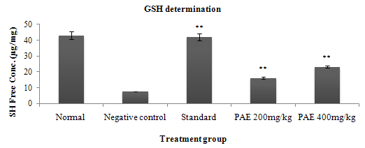
Fig. 1: Bar chart of GSH determination in Ethanol induced ulcer
The GSH result (Table-l) showed that minimal level of GSH was present in negative control group, indicated toxicity of Ethanol at cellular level by deduction of free SH concentration. Protective effect of both drug was present up to a significant level because P<0.05 when compared to the all group against negative control treated group. Prunus amygdalus extract did not show dose depended manner due to presence of P>0.05 when compare to both dose group (200mg/kg, 400mg/kg) of Prunus amygdalus.
GWM determination
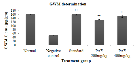
Fig. 2: Bar chart of GWM determination in Ethanol induced ulcer
According data of Table l concluded that among all groups level of gastric mucosa of minimum in negative control treated group and maximum was in ranitidine group. Drug treated groups had a significant difference (P<0.05) when compare to the negative control and standard group that means all group protects the mucosa but not up to standard group.
Hexosamine Determination
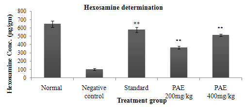
Fig. 3: Bar chart of Hexosamine determination in Ethanol induced ulcer
Hexosamine are precursor of biochemical synthesis of glycosylated protein and lipids. Both glycosylated and lipid are basic component of cell membrane and responsible for cell rigidity. In this present study was showed that there was present a significant protection for Hexosamine in all treated groups in compare to negative controlgroup.
Catalase Determination
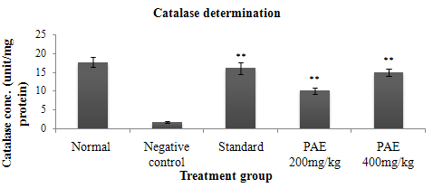
Fig. 4: Bar chart of Catalase determination in Ethanol induced ulcer
NOW YOU CAN ALSO PUBLISH YOUR ARTICLE ONLINE.
SUBMIT YOUR ARTICLE/PROJECT AT articles@pharmatutor.org
Subscribe to Pharmatutor Alerts by Email
FIND OUT MORE ARTICLES AT OUR DATABASE
Above data of Table l showed that catalase concentration per mg protein was higher in drug and ranitidine group with significant difference (P<0.05) in compare to vehicle group means least protection was present in negative control group. Dose depended manner was absent in drug treated group due to non significant level (P>0.05) between both doses of drug treated groups.
LPO determination
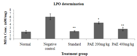
Fig. 5: Bar chart of LPO determination in ethanol induced ulcer
The LPO results in (Table l) showed that there was significance level difference present in negative control group and all other group that indicated that lipid peroxides enzyme was higher in vehicle treated group. Both extacts was reduced LPO at significant level and there was present dose depended manner because no significant level (P>0.05) was there between 200mg/kg and 400mg/kg treated group.
Ulcer Score
|
Treatment Group |
Ulcer Score of stomach in Ethanol induced ulcer |
|
Negative Control |
4.66±0.2108 |
|
Standard |
1.833±0.3073** |
|
PAE (200 mg/kg) |
2.833±0.3073* |
|
PAE (400 mg/kg) |
2.333±0.4216** |
Table-2: Observation of ulcer score in Ethanol induced ulcer
Each group consists of six animals, Data is presented in mean ±SEM
Here * means no significant difference (p > 0.05) in compare to negative control group
** means significant difference (p < 0.05) in compare to negative control group
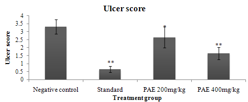
Fig. 6: Bar chart of ulcer score in Ethanol induced ulcer
Ulcer score is the counting of spots and severity of damage in gastric part by any moiety such as ethanol. This present study showed that all drug groups showed protection against ethanol damage by significant level (P<0.05) in compare to negative control group. Single drug treatment (200 mg/kg and 400mg/kg of P.A.E.) was effective up to a significant level (P<0.05) in compare to negative control group and also showed that drugs did not have dose depended manner because there was none significant level was resent between 200mg/kg/and 400mg/kg dose treatment of P.A.E.
Morphology of Stomach in Ethanol Induced Ulcer
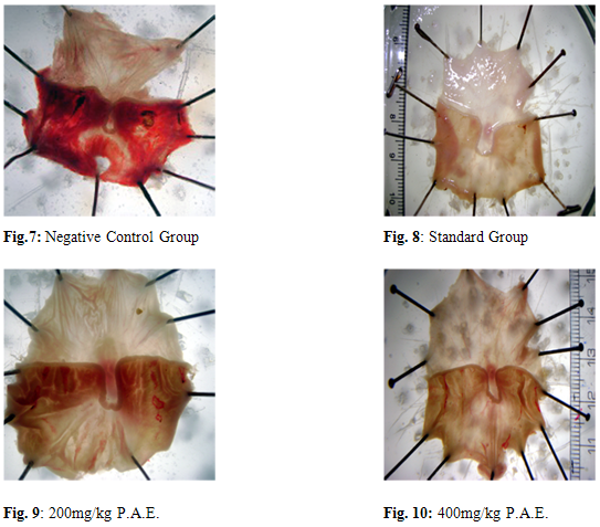
Result & Discussion
In the present study, herbal treated groups showed significant decrease in ulcer index as compared to control animals. Further, no significant difference was observed between herbal combination treated animals and ranitidine treated animals which indicate that the gastro protective effect caused by herbal treated group is comparable to ranitidine. It is reported that proton pump inhibitor such as lansoprazole and prostaglandin analogues help in reducing the risk of ulcer colitis specially bleeding type.
The level of GMW and HEXOSAMINE (in ethanol induced) was significantly decreased in negative control group as compared to normal group. Administration of herbal extract (200 mg/kg & 400mg/kg p.o.) increased GMW and HEXOSAMINE levels when compared to the disease control animals, which suggests its efficacy in preventing mucous damage. However, significant increase was observed all doses but in extract 400mg/kg were better results.
The level of CAT and GSH was significantly decreased in diseased group as compared to normal group. Administration of Prunus amygdalus (200 mg/kg & 400mg/kg p.o.) increased CAT and GSH levels as compared to the negative control animals, which suggests its efficacy in preventing free radical-induced damage. However, significant increase was observed in all doses but in 400mg/kg shows better result.
Lipid peroxidation is a free radical mediated process, which has been implicated in a variety of disease states such as hepatotoxicity, atherosclerosis, hypertension etc. lipid peroxidation mediated by free radicals is believed to be an important cause of destruction of and damage to cell membrane, which has been demonstrated to play an important role in pathogenesis of gastric mucosal injury induced by ethanol. Lipid peroxidation product, malondialdehyde (MDA) was found to be significantly increased in diseased group as compared to normal group. Our study has revealed a significant decrease in lipid peroxidation by herbal extract at all the doses under investigation.
Most widely used method for producing experimental gastric ulcers is pylorus ligation, as it is suitable for first line antiulcer screening; because the agents are retained in the stomach and may act by a variety of mechanisms. Reproducibility and high incidence of ulceration has been reported by this method. A major advantage of this method is that one can measure gastric secretory rate, percentage ulceration and ulcer severity in the same animal. It is reported that herbal extract significantly inhibited lesion formation in the glandular stomach and reduced acid secretory parameter and volume of gastric secretions.
Antacids such as aluminium hydroxide gel mainly act by decreasing pH of gastric juice and hence reduce total acidity of gastric juice. Anticholinergics such as pirenzipene inhibit effect of vagal stimulation i.e they compete with acetylcholine for M1 receptor on gastric parietal cell and hence decreases acid output. Proton pump inhibitors also acts by decreasing the gastric acid output. This mucus consists of mucin type of glycoprotein. This high molecular weight glycoprotein are mainly responsible for viscous and gel forming capacity of mucus. Increased mucus secretion by gastric mucousal cell prevent gastric ulceration by several mechanism such as lessing of stomach wall friction during peristalsis and gastric contraction, improving the buffering of gastric juice and by acting as an effective barrier to back diffusion of H+ ions.
REFERENCES
1. Kumars J, Asokan BR, Ramasuamy S, Anti-ulcer activity of polyherbal formulation. Pharmacologyonline 2009; 3: 419-23.
2. Tripathi KD. Essentials of medical pharmacology, Defination of peptic ulcer. Ed 5th, 587.
3. Borella LE, DiJoseph JF, Nabi Mir G, Cytoprotective and antiulcer activities of the antacid Magaldrate in the rat. Arzneim Forsch/Drug Res 1989; 39: 786–89.
4. Franzone JS, Cirillo R, Cravanzola C, Cytoprotective activity of deboxamet: A possible interference with prostaglandin and prostacyclin metabolism in rat gastric mucosa. Int J Tiss Reac 1988; 10: 149–58.
5. Ohkawa H, Oniwa N, Yagi K, Assay of lipid peroxides in animal tissues by thiobarbituric acid reaction. Anal Biochem 1979; 95: 351-58.
6. Sedlak J, Rudolf L, Estimation of total protein-bound and non-protein bound sulphydryl groups in tissues with Ellman's reagent. Anal Biochem, 1968; 25: 192-205.
7. Aebi H, Catalase: In Methods in enzymatic analysis, Academic Press: 1974; 674-84.
8. Muthusamy P, Suresh AJ, Balamurugan G, Antiulcer Activity of Azima Tetracantha: A Biochemical Study. Res J Pharm Tech, 2009; 2: 344-348.
9. Hawk P, Oser B, Summerson W, Gastric analysis: In Practical Physiological Chemistry, Toronto: Blakinston, 1954; 378–80.
NOW YOU CAN ALSO PUBLISH YOUR ARTICLE ONLINE.
SUBMIT YOUR ARTICLE/PROJECT AT articles@pharmatutor.org
Subscribe to Pharmatutor Alerts by Email
FIND OUT MORE ARTICLES AT OUR DATABASE


