

 About Authors:
1Hardik R. Patel*, 2Upanita C. Patel
1Industrial biotechnologist, 2Microbiologist
Anand, Gujarat, India.
*hardikigbt@gmail.com
INTRODUCTION:
Cryopreservation and lyophilization of plant germplasm has obvious advantages over in vitro storage in term of space saving and improved phytosanitation. We compared cryopreserved and lyophilized leaf as sources for genomic DNA isolation by CTAB protocol and PVP protocol.Our results showed that cryopreservation of leaf tissue yielded high molecular weight genomic DNA. The DNA was suitable for restriction-enzyme digestion and as a template for polymerase chain reaction (PCR) amplification. While these results rule out cryopreserved tissue as a source for DNA isolation, the ability to freeze-dry, powder, and efficiently store voluminous tissue samples for later use in DNA and protein isolation could be of great benefit to laboratories involved in molecular genetics and molecular biology.
[adsense:336x280:8701650588]
Reference Id: PHARMATUTOR-ART-1587
Objective: To improve and apply advanced techniques for plant genetic resources conservation through cryopreservation and lyophilization
To optimize a protocol for cryopreservation and lyophilization of following plants.
o Terminalia arjuna
o Terminalia catappa
o Terminalia chebula
o Jatropha curcas
o Jatropha gossypifolia
To optimize a protocol for DNA extraction from said plants after cryopreservation and lyophilization
Taxonomical classification of plant species and its description
Family: Combretaceae and Euphorbiaceae
Combretaceae are trees, shrubs, and lianas comprising about 20 genera and 600 species. The leaves are simple, alternate or opposite, entire, stipules small or absent. The flowers are bisexual or sometimes unisexual, usually actinomorphic. The perianth arises from near the summit of a tubular epigynous zone; calyx of usually 4 or 5 distinct to slightly connate sepals; corolla commonly of 4 or 5 distinct petals, occasionally absent. The androecium of 4-10 stamens is adnate to the epigynous zone, commonly in two whorls, often strongly exserted. The gynoecium is a single compound pistil of 2-5 carpels; style and stigma 1; ovary inferior, with 1 locule containing 2-6 apical ovules pendulous on long funiculi. The nectary is usually a disk (often hairy) above the ovary. The fruit is 1-seeded, often a flattened, ribbed, or winged drupe [Lim T, 2012].
[adsense:468x15:2204050025]
Euphorbiaceae is a spurge family of flowering plants, in the order Malpighiales, containing some 7,500 species in 275 genera. The family consists of annual and perennial herbs and woody shrubs or trees, rarely climbers. Flowers are of one sex, with male and female flowers usually borne on the same plant. Petals are rarely present. Flowers of Euphorbia are in cup-shaped clusters called Cynthia, each of which seems to be a single female flower, consisting of a single pistil surrounded by several male flowers, each of which has a single stamen. These clusters of reduced flowers are enclosed by involucres (whorl) of bracts (modified leaves) that resemble a corolla, or whorl of flower petals.
Male flowers of the other genera have one to many stamens, free or joined. Female flowers have three-chambered ovaries that are superior. There are as many styles as there are ovary cavities. The fruit is a three-chambered capsule. Leaves are usually simple and are alternate (or rarely opposite or whorled) in arrangement along the stems. The stems of many species contain milky latex [Tripathi S and Srivastava M, 2008].
A. Terminalia arjuna
Terminalia arjunais a medicinal plant of the genus Terminalia. The plantis about 20-25 metres tall; usually has a buttressed trunk, and forms a wide canopy at the crown, from which branches drop downwards. It has oblong, conical leaves which are green on the top and brown below; smooth, grey bark; it has pale yellow flowers which appear between March and June; its glabrous, 2.5 to 5 cm fibrous woody fruit, divided into five wings, appears between September and November [Biswas M et al., 2011].
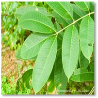
Figure 1:Terminalia arjuna
· Binomial name Terminalia arjuna
· Scientific classification [Nema R et al., 2012]
Kingdom: Plantae
Division: Magnoliophyta
Class: Magnoliopsida
Order: Myrtales
Family: Combretaceae
Genus: Terminalia
Species: T. arjuna
· Geographical location
Terminalia arjunais usually found growing on river banks or near dry river beds in West Bengal and South and Central India. Terminalia arjunais common throughout India especially in the sub Himalayan tracts and Eastern India. They are widely grown in Bandhavgarh National Park, Pench Tiger Reserve and Kanha National Park in India [Biswas M et al., 2011].
· Chemical constituents and components
Terminalia arjunaconsists of various components such as triterpenoids, tannins, flavonoids, oligomeric proanthocyanidins and saponin glycosides [Nema R et al., 2012].
· Medicinal uses
Terminalia arjunais used for the treatment of coronary artery disease, heart failure, edema, angina, hypertension and hypercholesterolemia [Biswas M, et al., 2011]. Its bark possesses diuretic, prostaglandin enhancing and coronary risk factor modulating properties. It is also considered as beneficial in the treatment of asthma [Devi R et al., 2007].
B. Terminalia catappa
Terminalia catappa is a large tropical tree in the Leadwood tree family, Combretaceae. It has corky, light fruit that is dispersed by water. The seed within the fruit is edible when fully ripe, tasting almost like almond. It grows to 35 metres (115 ft) tall, with an upright, symmetrical crown and horizontal branches. As the tree gets older, its crown becomes more flattened to form a spreading, vase shape. Its branches are distinctively arranged. The leaves are large, 15–25 centimeters (5.9–9.8 in) long and 10–14 centimeters (3.9–5.5 in) broad, ovoid, glossy dark green and leathery. The trees are monoecious, with distinct male and female flowers on the same tree. Both are 1 centimeter (0.39 in) in diameter, white to greenish, inconspicuous with no petals; they are produced on axillaries or terminal spikes. The fruit is a drupe 5–7 centimeters (2.0–2.8 in) long and 3–5.5 centimeters (1.2–2.2 in) broad, green at first, then yellow and finally red when ripe, containing a single seed [Hnawia E et al., 2011].

Figure 2: Terminalia catappa
· Binomial name Terminalia catappa
· Scientific classification [Hnawia E et al., 2011]
Kingdom:Plantae
Division: Magnoliophyta
Class: Magnoliopsida
Order: Myrtales
Family: Combretaceae
Genus: Terminalia
Species: T. catappa
· Geographical location
The tree has been spread widely by humans and the native range is uncertain. It has long been naturalised in a broad belt extending from Africa to Northern Australia and New Guinea through Southeast Asia and Micronesia, in subtropical and tropical zones of Indian Subcontinent and Pacific Oceans. It has also been planted extensively throughout the tropics. Subtropical and tropical maritime climates with annual rainfall generally 1000–3500 mm (40–140 in); elevations below 300–400 m (1000–1300 ft)It is distributed in the forest of Western UP and Haryana [Thomson L and Evans B, 2006].
· Chemical constituents and components
The leaves contain several flavonoids (like kaempferol or quercetin), several tannins (such as punicalin, punicalagin or tercatin), saponines and phytosterols [Hnawia E et al., 2011].
· Medicinal uses
Astringent in thrush, antidisenteric, mild diuretic, leaf-antiseptic, anti-inflammatory, diaphoretic, antiindigestion, antidysentery, for treating mouth infections, headache, colic [Deharo E et al., 2011].
C.Terminalia chebula
It is belongs to the family combretaceae. It is commonly called as Black myrobalan. It is a deciduous tree, younger stems glabrescent, woody. Leaves are 10 – 20 cm long, sub – opposite, simple; estipulate; petiolate; laminae broadly elliptic to elliptic – oblong, rarely ovate, the bases obtuse, the margins entire, the tips acute, glabrescent. Flowers are 2 mm long , 3-4 mm in diameter; bracts nearly glabrous, 1.5-2.0 mm long; calyx outside glabrous, inside densely villous, calyx-segments triangular; stamens 3-4 mm long; ovary glabrous, ovoid, 1 mm long; style glabrous, 2.5- 3.0 mm long. Seed is rough, ellipsoid, 1.0-2.0 cm by 0.2 -0.7 cm and without ridges [SuryaPrakash D et al., 2012;].

Figure 3: Terminalia chebula
· Binomial name Terminalia chebula
· Scientific classification
Kingdom: Plantae
Division: Magnoliophyta
Class: Magnoliopsida
Order: Myrtales
Family: Combretaceae
Genus: Terminalia
Species: T. chebula
NOW YOU CAN ALSO PUBLISH YOUR ARTICLE ONLINE.
SUBMIT YOUR ARTICLE/PROJECT AT articles@pharmatutor.org
Subscribe to Pharmatutor Alerts by Email
FIND OUT MORE ARTICLES AT OUR DATABASE
· Chemical constituents and components
Terminalia chebula contains the triterpenes arjun glucoside 1, arjungenin and the chebulosides 1&2. Other constituents contains tannins up to 30%, chebulic acid 3-5%, chebulinic acid 30%, tannic acid 20-40%, ellagic acid, 2,4-chebulyi–β-D-gluco pyranose, gallic acid, ethyl gallate, punicalagin terflavin A , terchebin, some purgative of the nature of anthraquinone , flavonoids like luteolin, rutins, and quercetin etc [SuryaPrakash D et al., 2012].
· Geographical location
T. chebula occurs scattered in teak forest, mixed deciduous forest, extending into forests of comparatively dry types. It grows in India, Myanmar, Bangladesh, Iran, Egypt, Turkey, China etc. In India Haritakitree is grows in deciduous forests and found in North India and South words to the Deccan table lands at 1000 to 3000 ft. In Myanmar country grows up to 5000 ft Terminalia chebula plant is a native plant in India and Southeast Asia and is extensively cultivated in Taiwan. [SuryaPrakash D et al., 2012;Said M et al., 2012].
· Medicinal uses
It is used to increase the appetite, as digestive aid liver stimulant, as stomachic, as gastro-intestinal prokinetic agent and as mild laxative. It is indicated in protracted diarrhea with hematochezia and prolapse of rectum. It is a good nervine, used in nervous weakness, nervous irritability.It is helpful in renal calculi, dysurea, and retention of urine and used for treating parasitic infection. It is used as a blood purifier, gargle for sore throat, ulcerated gums, and muscular rheumatism [SuryaPrakash D et al., 2012].
D. Jatropha gossypifolia
It is belongs to the family Euphorbiaceae and the order, “Geraniale”.The common name for J. gossypifolia is bellyache bush, pignut or fignut, and in Yoruba land it is commonly known as “Lapalapa”. It is a bushy, gregarious shrub, up to 1.8m, 3-5 lobed, approximately 20 cm long and wide, with leaves having a long petiole, covered with glandular hairs. The seed are greenish capsule-like seeds. The leaf stalks are covered with coarse dark brown hairs and the young leaves are sticky. It has thin, often greenish bark, which exudes copious amount of watery sap when cut. The fruits are three-celled with one seed per cell. J. gossypifolia is widely cultivated as ornamental plant. It is the common red species planted around houses. It is also planted to control the soil erosion along the slopes. It prefers arid environment [Seth R and Sarin R, 2010].

Figure 4: Jatropha gossypifolia
· Binomial name Jatropha gossypiifolia
· Scientific classification
Kingdom: Plantae
Order: Geraniale
Family: Euphorbiaceae
Genus: Jatropha
Species: J. gossypiifolia
· Chemical constituents and components
The major phytochemicals of interest are alkaloids, tannins, flavonoids, phenolic compounds, and steroidal saponins; however, other diverse groups of naturally occurring phytochemicals such as unsaturated sterols, triterpenoids, and essential oils are also present [Seth R and Sarin R, 2010].
• Geographical location
Native to tropical America, but is now cultivated widely in tropical countries throughout the world. It is grown occasionally in warmer parts of Australia and is naturalised in a few places in Queensland and the Northern Territory. In Florida it is found chiefly south of Orlando. It is also a common plant in the Hawaiian Islands. Introduced to southern Africa, the plant has spread from Mozambique through Zambia to the Transvaal and Natal. This species is also found throughout the warmer parts of Asia. This plant is normalized in many parts of India. It has also naturalized in most tropical area of the world [Jadhav A et al., 2012].
· Medicinal uses
The leaves of J. gossypifolia are used for intermittent fevers, carbuncles, eczema, itches, sores on the tongues of babies, swollen mammae, stomachache, and venereal disease. The leaf decoction is used for bathing wounds [Oduola T et al., 2007].
E. Jatropha curcas
Jatropha curcas L belongs to the euphorbia family. It is a large coarse annual shrub or small short lived tree which can grow 3.5 to 4.5 metres (8-15 feet) tall. It has thin, often greenish bark which exudes copious amounts of watery sap when cut. Leaves are dark green; alternate, simple, ovate to slightly lobed with 3-5 indentations. Petioles are 10cm (4 inches) long. Flowers are yellow to green in color, borne in axils of the leaves and being small is mostly hidden by foliage. Fruit are small capsule-like, round fruit; about 2.5 - 4 cm (1-1.5 inches) in diameter. These are green and fleshy when immature, becoming dark brown when ripe and splitting to release 2 or 3 black seeds each about 2 cm (3/4 inch) long. The meat of the seeds is white and oily in texture and is reported to have an agreeable taste. [inchem.org/documents/pims/plant/jcurc.htm]

Figure 5: Jatropha curcas
· Binomial name: Jatropha curcas
· Scientific classification
Kingdom: Plantae
Order: Malpighiales
Family: Euphorbiaceae
Genus: Jatropha
Species: J. curcas
· Chemical constituents and components
The phytochemicals analysis of the extract revealed the presence of triterpenoids, volatile oils, alkaloids, flavonoids, saponins and tannins [Uche F and Aprioku J, 2008].
• Geographical location
It is widely cultivated as an ornamental. Native to tropical America, but is now cultivated widely in tropical countries throughout the world. It is grown occasionally in warmer parts of Australia and is naturalised in a few places in Queensland and the Northern Territory. It has been planted in Gujarat in Ahmedabad, Kutch, Bhavnagar, Jamnagar, Surat, Banaskantha, Rajkot, and Surendranagar. It is one of the 18 species found scattered in various parts of India [Punia M, 2007].
· Medicinal uses
Most parts of this plant are used for the treatment of various human and veterinary ailments. The white latex serves as a disinfectant in mouth infections in children.The leaves contain apigenin, vitexin and isovitexin etc. which along with other factors enable them to be used against malaria, rheumatic and muscular pains [Thomas R et al., 2008].
METHODOLOGY
COLLECTION OF SAMPLE
Total 5 plant species were collected for herbarium preparation as well as DNA isolation from Indroda Botanical Garden, Gandhinagar [Figure 4.3.1]. For herbarium preparation the plant specimen was placed on a filter paper. The leaves were arranged to avoid overlapping and the respective filter papers were kept in the herbarium press.
Table 1: List of plant species
|
Plants name |
Family |
Latitude |
Longitude |
|
Terminalia arjuna |
Combretaceae |
N 23° 11.650 |
E 72° 38.853 |
|
Terminalia catappa |
Combretaceae |
N 23° 11.659 |
E 72° 38.861 |
|
Terminalia chebula |
Combretaceae |
N 23° 11.722 |
E 72° 38.936 |
|
Jatropha curcas |
Euphorbiaceae |
N 23° 11.665 |
E 72° 38.809 |
|
Jatropha gossypifolia |
Euphorbiaceae |
N 23° 11.664 |
E 72° 38.809 |
HERBARIUM PREPARATION
1. The plant sample which was earlier collected from the field was taken out from the herbarium press.
2. The plant samples were treated with mercuric chloride solution.
3. The treated plant samples were placed in a new blotting paper and name of the plant, date of collection and name of the collector were written.
4. All the other plant samples were treated similarly and put in the herbarium press.
5. The blotting paper was changed every alternate day till the plant samples were dried completely.
6. After the samples were dried, they were taken out from the herbarium press and stuck on the herbarium sheet with the help of fevicol.
7. The details about the plant were filled on the herbarium sheet.
CRYOPRESERVATION
Surface sterilization
Leaves were sterilized by surface washing under tap water than distilled water to remove dust and other contaminants.
Weighing of sample
The leaves were dried with blotting paper and approximately 20g leaves were weighed without middle ribs.
Sample preparation for cryopreservation
Leaves were crushed into liquid nitrogen to make a fine powder and 3 cryovials were filled up for each plant and were cryopreserved at -196°C cryopreservation was carried out by placing the cryovials in canisters and immersing them in the cryocan containing liquid nitrogen. DNA extraction was carried out from cryopreserved sample at intervals of 1, 7 and 15 days.
LYOPHILIZATION
Surface sterilization
Leaves were sterilized by surface washing under tap water than distilled water to remove dust and other contaminants.
Sample preparation for lyophilization
The leaves were dried with blotting paper and middle ribs were removed. They were crushed into liquid nitrogen to make fine powder and approximately 15g were weighed. After lyophilization, the powder was collected in petridish which were sealed with cling film.
Lyophilization process
Plant samples were lyophilized at vacuum of 0.16 mbar and -500C for 72 hrs. Lyophilized powder was stored in tightly sealed petridish at room temperature.
The Super Modulo freeze dryer used for lyophilization at GSBTM does not facilitate the temperature and pressure regulation. The protocol for operation of this instrument was as follows:
1. The powdered samples were frozen for some time at -20°C freezer.
2. The frozen leaves were evenly spread on the product trays without the delay for the leaves to cool down again.
3. The racks with the samples were placed in the dome and covered with the acrylic lid.
4. It was made sure that all the detachable parts associated with vacuum are well greased so as to minimize the chances of any leakage.
5. The “Fridge” switch was turned on. Within 1 hr, the chamber temperature should be around -40°C to -50°C.
6. The “pump” switch was turned on. The pressure reached up to 10-1 mbar.
7. The samples were lyophilized for 1 whole day.
8. After the run was complete, the “Pump” switch was turned off. The knobs were gradually opened to release the pressure.
9. The samples were placed in a dry, air tight environment as soon as possible and stored out of direct sunlight [Super Modulo Instruction Manual, 2006].
Residual moisture content:
The moisture content was determined using following formula.
Moisture content (%) = (fresh weight – dry weight) ×100
fresh weight
Residual moisture content (%) = 100- Moisture content
DNA EXTRACTION
DNA extraction was carried out from cryopreserved, lyophilized and oven dried plant samples using two different protocols which are mentioned below as follows:
A. CTAB protocol [Doyle J and Doyle J, 1987]
- Water bath was set to 65 ºC and CTAB buffer was put into the water bath.
- 100 mg of plant material (cryopreserved, lyophilized, oven dried samples) was taken in a motor pestle.
- Liquid nitrogen was added and the sample was crushed to powder form. (In case of already crushed samples, proceed to next step)
- 3 ml of 3% CTAB buffer, containing 6% PVP (pH 8) and 6.1 µl of β-mercaptoethanol was added.
- The solution was incubated at 65ºC for 1 hour.
- The tube was centrifuged at 12,000 rpm for 5’ at 4 0C by using centrifuge (Eppendorf).
- The supernatant was collected in fresh tube and 5µl/ml of Proteinase K was added and incubated at 65 °C for 1 hr.
- Equal volume of P: C: I was added, mixed gently, centrifuged 12000 rpm for 15 mins.
- Supernatant was collected, equal volume of C: I was added, mixed and centrifuged at 12000 rpm for 15 mins.
- Step 9 was repeated.
- 5 µl of 20 mg/ml RNase A or 10µl of 10 mg/ml RNase A was added, incubated at 65 °C for 1 hour or 37 °C overnight.
- 1/10th volume of 3M sodium acetate and equal volume of absolute ethanol was added, incubated for 20 mins at -20°C.
- The samples were centrifuged at 12000 rpm for 10 mins, supernatant was discarded and the pellet was recovered.
- The pellet was washed twice with 70% ethanol and centrifuged at 10000 rpm for 10 mins.
- The pellet was allowed to air dry and dissolved in 200 µl of 1X TE buffer.
NOW YOU CAN ALSO PUBLISH YOUR ARTICLE ONLINE.
SUBMIT YOUR ARTICLE/PROJECT AT articles@pharmatutor.org
Subscribe to Pharmatutor Alerts by Email
FIND OUT MORE ARTICLES AT OUR DATABASE
PVP protocol [KIM Cet al., 1997]
- Approximately 100 mg of sample was ground in a pestle in 5 µl (one drop) of 1% (v/v) 2-mercaptoethanol.
- After grinding, 300 µl of extraction was added to the homogenate, which was transferred to a 1.5 ml tube and the tube was flicked at the bottom occasionally to keep the extract mixed.
- The homogenate was incubated at room temperature for 1 hr.
- Freshly prepared PVP (6% of final volume) and one half volume of 7.5 M ammonium acetate were added separately.
- The mixture was incubated on ice for 30 min and centrifuged for 10 min in a micro centrifuge (10 000 g at 4 °C).
- The supernatant was transferred to a fresh tube to which was added 1 volume of isopropanol, and left at -20 °C for 30 min to precipitate the DNA.
- After centrifugation at 10,000 g for 10 min, the supernatant was discarded and the DNA pellet was air-dried.
- The DNA pellet was resuspended in 500 µl TE buffer (10 mM Tris–HCl pH 8.0, 0.1 mM EDTA pH 8.0) or distilled water.
- Two µl of RNase (1 mg/ml) was added to the solution and incubated at 37°C for 15min.
- One volume chloroform: isoamyl alcohol (24:1) was added and emulsified by inverted shaking to remove both RNase and plant pigments.
- The above step was repeated once again.
- After centrifugation (10,000 g at 4 °C) for 5 min, the supernatant was transferred to a fresh tube to which 1 volume of isopropanol was added and left at –20 °C for 10 min.
- After centrifugation at 10,000 g for 10 min, the pellet was washed with 1 ml 80% ethanol and air-dried.
- The DNA pellet was re-dissolved in 30 µl TE or distilled water.
AGAROSE GEL ELECTROPHORESIS
Agarose gel electrophoresis was carried out to check for the presence of genomic DNA isolated from plant samples.
Method
1. The required amount of agarose was weighed, 0.8% agarose was prepared in 0.5X TBE buffer. After heating the solution and letting it to cool, required amount of ethidium bromide was added.
2. The tank was filled up with 0.5X TBE buffer and the gel was immersed in it.
3. 5 µl of sample with 2 µl of bromophenol blue was mixed and the samples were loaded in the respective lanes.
4. The gel was run at 80-100 V for 1 hour.
5. The agarose gel was observed under the Gel-Documentation system (Bio-Rad).
6. The image was captured, required lanes were labeled and the image was saved.
RESULTS AND DISCUSSION
High quality of DNA, RNA and protein are required for meaningful molecular biology studies. Several extraction methods have recently been developed for DNA, RNA and protein extraction. However, many of these techniques require either fresh tissue or tissue stored and strictly maintained at ultralow low temperatures at -50 ?C to -70 ?C [Saha S et al., 1997]. At this ultra-low temperature, all cellular divisions and metabolic processes are stopped, allowing conservation for a theoretically unlimited period of time [Engelmann, 2004]. Generally, only a limited number of freshly collected tissue samples can be extracted at any given time, while ultralow freezer space for storage of bulky tissue samples is usually at a premium in most laboratories. A solution to this problem is lyophilization of the plant tissues by grinding them as dry powder for efficient storage in limited freezer space [Saha S et al., 1997].
In this study, we had tried optimizing a protocol for lyophilization, cryopreservation and DNA extraction of five socio-economically and medically important plant species. We had also tried to study whether the lyophilized plant leaves were suitable as a source for DNA isolation. A comparative analysis was also performed to find out which among cryopreservation or lyophilization was a better method for storage of plant leaf tissues, by isolation of DNA from the cryopreserved and lyophilized leaves. Three plant species, Terminalia arjuna, Terminalia catappa andTerminalia chebula belonging to Combretaceae family and two plant species Jatropha curcas and Jatropha gossypifolia, belonging to Euphorbiaceae family were selected for the study. The selected plant species have medicinal value which can be used for the treatment of human disease and diagnosis.
For optimization of DNA extraction method, two protocols were performed one of which was a modified CTAB protocol [Doyle J and Doyle J, 1987] and the other was PVP protocol [Kim C et al., 1997]. The CTAB protocol gave better results than PVP protocol for DNA isolation from cryopreserved leaf samples while PVP protocol gave better results only in case of lyophilized leaves.
In case of lyophilization, the concentration and the purity (A260/A280) of the DNA extracted was in the range of 887 ng/µl - 1200 ng/µl and 1.130 - 1.334 respectively. The purity and concentration of Jatropha curcas was found after storage of the lyophilized plant leaves for up to 15 days [Table 2]. The residual moisture content post lyophilization was as low as 22.8 % in Terminalia catappa as high as 35.06 % in Terminalia chebula [Table 3, Figure 5.6]. The gel images are shown in Figure 5.8.
In case of cryopreservation, the concentration and the purity of the DNA extracted was in the range of 40 ng/µl - 1642 ng/µl and 0.917 – 1.778 respectively. Bands of DNA were observed in cryopreserved leaf samples of Terminalia chebula stored for up to 15 days and for the remaining four species for up to 7 days [Table 4, Figure 5.7].
Many factors affect the preservation of DNA, including the type of plant, the chemical and physical environment in which that plant is stored, and the duration of storage and the preservation method. However, the interactions of these factors and their resultant effects on DNA preservation are difficult to predict [Dawson M et al., 1998] and the protocol for DNA extraction needs to be further optimized. The ratio of purity of DNA was found to be less than 1.8 with high concentration in lyophilized samples of Terminalia arjuna, Terminalia catappa, Terminalia chebula; therefore it indicates the presence of other impurities like polyphenols, polysaccharides, etc.
The purity of the DNA extracted from both cryopreserved and lyophilized leaves was in the range of 0.917 – 1.778. A ratio less than 1.8 indicates the probable presence of proteins and/or UV absorbers and a ratio higher than 2.0 indicates that the sample may be contaminated with chloroform or phenol [Reddy J, 2009].
Table 2: Concentration and purity of the genomic DNA extracted by CTAB and PVP protocol after preservation of the sample through lyophilization
|
Sr. No. |
Name of plant species |
Residual moisture content (%) |
Protocol for DNA extraction |
Days of storage |
Purity of the DNA extracted (A260 |
Conce |
|
1. |
Terminalia arjuna |
30.2667
|
Protocol 1(CTAB) |
1 |
0.986 |
365 |
|
7 |
1.677 |
110 |
||||
|
15 |
1.073 |
220 |
||||
|
Protocol 2 |
15 |
1.187 |
555 |
|||
|
2. |
Terminalia catappa |
22.8
|
Protocol 1(CTAB) |
1 |
1.412 |
85 |
|
7 |
1.64 |
60 |
||||
|
15 |
1.026 |
200 |
||||
|
Protocol 2 |
15 |
1.474 |
140 |
|||
|
3. |
Terminalia chebula |
35.0667
|
Protocol 1(CTAB) |
1 |
1.158 |
293 |
|
7 |
1.5 |
387 |
||||
|
15 |
1.049 |
160 |
||||
|
Protocol 2 |
15 |
1.737 |
82.5 |
|||
|
4. |
Jatropha curcas |
23.7333
|
Protocol 1(CTAB) |
1 |
1.934 |
885 |
|
7 |
1.655 |
300 |
||||
|
15 |
1.176 |
100 |
||||
|
Protocol 2 |


