

 About Authors:
P.Ramanujaiah*, Dr M.Purushothaman, Hemalatha
Vasavi institute of pharmaceutical sciences, Kadapa
*rama.mpharm@gmail.com
Abstract:
The application of nanotechnology to drug delivery has currently more than 20 Nanoparticles therapeutics are in clinical use, validating the ability of Nanoparticles to improve the therapeutic index of drugs. In addition to the already approved Nanoparticles, numerous other Nanoparticles platforms are currently under various stages of preclinical and clinical development, including various liposomes, polymeric micelles, dendrimers, quantum dots, gold Nanoparticles, and ceramic Nanoparticles. With continued research and development efforts, nanotechnology is expected to have a tremendous impact on medicine for decades to come.
[adsense:336x280:8701650588]
Reference Id: PHARMATUTOR-ART-1523
Introduction:
Nanotechnology refers to the use man-made of nano-sized particles for industrial or medical applications suited to their unique properties. Physical properties of known elements and materials can change as their surface to area ratio is dramatically increased, i.e. when nanoscale sizes are achieved. These changes do not take place when going from macro to micro scale. Changes in physical properties such as colloidal properties, solubility and catalytic capacity have been found very useful in areas of biotechnology, such as bioremediation and drug delivery. The very different properties of the different types of nanoparticles have resulted in novel applications. The extremely small size of nanoparticles allows them to penetrate cells and interact with cellular molecules. Nanoparticles often also have unique electrical properties and make excellent semiconductors and imaging agents.
Nanotechnology is the understanding and control of matter generally in the 1–100nm dimension range. The application of nanotechnology to medicine, known as nanomedicine, concerns the use of precisely engineered materials at this length scale to develop novel therapeutic and diagnostic modalities.Nano materials have unique physicochemical properties, such as ultra small size, large surface area to mass ratio, and high reactivity, which are different from bulk materials of the same composition. These properties can be used to overcome some of the limitations found in traditional therapeutic and diagnostic agents.
The use of materials in nanoscale provides unparallel freedom to modify fundamental properties such as solubility, diffusivity, blood circulation half-life, drug release characteristics, and immunogenicity. In the last two decades, a number of Nanoparticles-based therapeutic and diagnostic agents have been developed for the treatment of cancer, diabetes, pain, asthma, allergy, infections, and so on these nanoscale agents may provide more effective and/or more convenient routes of administration, lower therapeutic toxicity, extend the product life cycle, and ultimately reduce health-care costs. As therapeutic delivery systems, Nanoparticles allow targeted delivery and controlled release. For diagnostic applications, Nanoparticles allow detection on the molecular scale: they help identify abnormalities such as fragments of viruses, precancerous cells, and disease markers that cannot be detected with traditional diagnostics. Nanoparticles-based imaging contrast agents have also been shown to improve the sensitivity and specificity of magnetic resonance imaging. Given the vast scope of nanomedicine, we will focus on the therapeutic applications, in particular, drug delivery applications, of Nanoparticles.
Many advantages of Nanoparticles-based drug delivery have been recognized.It improves the solubility of poorly water-soluble drugs, prolongs the half-life of drug systemic circulation by reducing immunogenicity, releases drugs at a sustained rate or in an environmentally responsive manner and thus lowers the frequency of administration, delivers drugs in a target manner to minimize systemic side effects, and delivers two or more drugs simultaneously for combination therapy to generate a synergistic effect and suppress drug resistance. As a result, a few pioneering Nanoparticles-based therapeutic products have been introduced into the pharmaceutical market and numerous ensuing products are currently under clinical testing or are entering the pipeline.
Out of plethora of size-dependant physical properties available to someone who is interested in the practical side of Nano materials, optical and magnetic effects are the most used for biological applications. Nanomedicine seeks to deliver a valuable set of research tools and clinically useful devices in the near future. The National Nanotechnology Initiative expects new commercial applications in the pharmaceutical industry that may include advanced drug delivery systems, new therapies, and in vivo imaging.
Neuro-electronic interfaces and other nanoelectronics-based sensors are another active goal of research. Further down the line, the speculative field of molecular nanotechnology believes that cell repair machines could revolutionize medicine and the medical field.
Important Characteristics of Nanomedicine:
* Target specificity
* Efficient drug release
* Extreme small size
* neuro-electronic interfaces
* Carry high concentrated drug
[adsense:468x15:2204050025]
Target specificity:
It is greatly observed that nanoparticles are promising tools for the advancement of drug delivery, medical imaging, and as diagnostic sensors. However, the biodistribution of these nanoparticles is still imperfect due to the complex host's reactions to nano- and microsized materials and the difficulty in targeting specific organs in the body. Nevertheless, a lot of work is still ongoing to optimize and better understand the potential and limitations of nanoparticulate systems.
For example: current research in the excretory systems of mice shows the ability of gold composites to selectively target certain organs based on their size and charge. These composites are encapsulated by a dendrimer and assigned a specific charge and size. Positively-charged gold nanoparticles were found to enter the kidneys while negatively-charged gold nanoparticles remained in the liver and spleen. It is suggested that the positive surface charge of the nanoparticle decreases the rate of opsonization of nanoparticles in the liver, thus affecting the excretory pathway.
Even at a relatively small size of 5 nm, though, these particles can become compartmentalized in the peripheral tissues, and will therefore accumulate in the body over time. While advancement of research proves that targeting and distribution can be augmented by nanoparticles, the dangers of nanotoxicity become an important next step in further understanding of their medical uses.
Efficient drug release:
The basic point to use drug delivery is based upon three facts:
* Efficient encapsulation of the drugs
* Successful delivery of said drugs to the targeted region of the body
* Successful release of that drug there.
Drug delivery systems, lipid- or polymer-based nanoparticles can be designed to improve the pharmacological and therapeutic properties of drugs. nanoparticles have beneficial properties that can be used to improve drug delivery. Where larger particles would have been cleared from the body, cells take up these nanoparticles because of their size. Complex drug delivery mechanisms are being developed, including the ability to get drugs through cell membranes and into cell cytoplasm.
Efficiency is important because many diseases depend upon processes within the cell and can only be impeded by drugs that make their way into the cell. Triggered response is one way for drug molecules to be used more efficiently. Drugs are placed in the body and only activate on encountering a particular signal. . For example, a drug with poor solubility will be replaced by a drug delivery system where both hydrophilic and hydrophobic environments exist, improving the solubility’s also, a drug may cause tissue damage, but with drug delivery, regulated drug release can eliminate the problem. If a drug is cleared too quickly from the body, this could force a patient to use high doses, but with drug delivery systems clearance can be reduced by altering the pharmacokinetics of the drug.
Poor bio-distribution is a problem that can affect normal tissues through widespread distribution, but the particulates from drug delivery systems lower the volume of distribution and reduce the effect on non-target tissue. Potential nanodrugs will work by very specific and well-understood mechanisms; one of the major impacts of nanotechnology and nanoscience will be in leading development of completely new drugs with more useful behavior and less side effects.
NOW YOU CAN ALSO PUBLISH YOUR ARTICLE ONLINE.
SUBMIT YOUR ARTICLE/PROJECT AT articles@pharmatutor.org
Subscribe to Pharmatutor Alerts by Email
FIND OUT MORE ARTICLES AT OUR DATABASE
Extreme small size:
The size of nanomaterials has great influence on their properties. The very different properties of the different types of nanoparticles have resulted in novel applications. The extremely small size of nanoparticles allows them to penetrate cells and interact with cellular molecules. Nanoparticles often also have unique electrical properties and make excellent semiconductors and imaging agents.
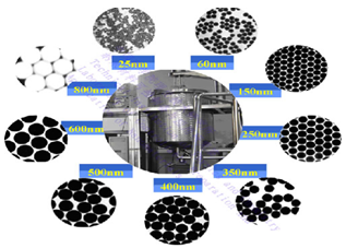
Fig: 1 Controlled Synthesis Silica Particles with Various Sizes
Fig: 2 Controlled Synthesis Gold colloids with Various Sizes
Fig: 3 Controlled Synthesis Metal colloid
Neuro-Electronic Interfaces:
Neuro-electronic interfacing is a visionary goal dealing with the construction of nanodevices that will permit computers to be joined and linked to the nervous system. This idea requires the building of a molecular structure that will permit control and detection of nerve impulses by an external computer. The computers will be able to interpret, register, and respond to signals the body gives off when it feels sensations. The demand for such structures is huge because many diseases involve the decay of the nervous system. Also, many injuries and accidents may impair the nervous system resulting in dysfunctional systems and paraplegia. If computers could control the nervous system through neuro-electronic interface, problems that impair the system could be controlled so that effects of diseases and injuries could be overcome.
Two considerations must be made when selecting the power source for such applications. They are refuelable and nonrefuelable strategies. A refuelable strategy implies energy is refilled continuously or periodically with external sonic, chemical, tethered, magnetic, or electrical sources. A nonrefuelable strategy implies that all power is drawn from internal energy storage which would stop when all energy is drained.
One limitation to this innovation is the fact that electrical interference is a possibility. Electric fields, electromagnetic pulses (EMP), and stray fields from other in-vivo electrical devices can all cause interference. Also, thick insulators are required to prevent electron leakage, and if high conductivity of the in-vivo medium occurs there is a risk of sudden power loss and “shorting out.” Finally, thick wires are also needed to conduct substantial power levels without overheating. Little practical progress has been made even though research is happening.
The wiring of the structure is extremely difficult because they must be positioned precisely in the nervous system so that it is able to monitor and respond to nervous signals.
The structures that will provide the interface must also be compatible with the body’s immune system so that they will remain unaffected in the body for a long time15 In addition, the structures must also sense ionic currents and be able to cause currents to flow backward. While the potential for these structures is amazing, there is no timetable for when they will be available.
APPLICATIONS:
The use of nanotechnology in medicine offers some exciting possibilities. Some techniques are only imagined, while others are at various stages of testing, or actually being used today. A list of some of the applications of nanomedicine to biology or medicine is given below.
* Molecular imaging therapy
* Prevention &control
* Early detection
* Diagnosis imaging
* Multifunctional
Molecular imaging therapy:
The small size of Nanoparticles endows them with properties that can be very useful in oncology, particularly in imaging. Quantum dots (Nanoparticles with quantum confinement properties, such as size-tunable light emission), when used in conjunction with MRI (magnetic resonance imaging), can produce exceptional images of tumor sites. These Nanoparticles are much brighter than organic dyes and only need one light source for excitation. This means that the use of fluorescent quantum dots could produce a higher contrast image and at a lower cost than todays organic dyes used as contrast media. The downside, however, is that quantum dots are usually made of quite toxic elements.
Another Nano property, high surface area to volume ratio, allows many functional groups to be attached to Nanoparticles, which can seek out and bind to certain tumor cells. Additionally, the small size of Nanoparticles (10 to 100 nanometers), allows them to preferentially accumulate at tumor sites. A very exciting research question is how to make these imaging Nanoparticles do more things for cancer. For instance, is it possible to manufacture multifunctional Nanoparticles that would detect, image, and then proceed to treat a tumor.
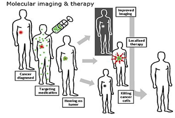
Fig: 4 molecular imaging and therapy
Sensor test chips containing thousands of nanowires, able to detect proteins and other biomarkers left behind by cancer cells, could enable the detection and diagnosis of cancer in the early stages from a few drops of a patient's blood.The basic point to use drug delivery is based upon three facts:
a) Efficient encapsulation of the drugs
b) Successful delivery of said drugs to the targeted region of the body.
c) Successful release of that drug.
Nanotechnology Used In Cancer Treatment:
They are smaller than human cells 10,000 to 20,000 nanometers in diameter and similar in size to large biological macromolecules such as enzymes and receptors. Nanostructures can be so small that the body may clear them too rapidly for them to be effective in detection or imaging. Larger nanoparticles may accumulate invital organs, creating a toxicity problem. Scientists will need to consider these factors as they attempt to create nanodevices the body will accept.
The goal of nanodevices:
* Recognize Precancerous Or Cancerous Cells.
* Release A Drug That Targets Only Cancer Cells.
* Report back On the Effectiveness Of the Treatment.
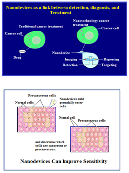
Fig: 5 sensitivity improving nano device
1. Photothermal Ablation:
* Cancer cells die at 42° C (108° F), normal cells die at about 46° C (115° F)
* Current optical fiber treatment
* Nanoparticles can be tuned to be Excited only by certain ranges of light
* Hollow, gold nanospheres are 50 times more effective at absorbing light near the infrared than solid gold nanoparticles
2. MRI Contrast Agents:
* Using a metal-organic framework with metals such as gadolinium.
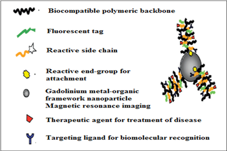
Fig: 6 different types of attachments
3. Fluorescent Microscopy:
* these are used in detection of cancer cells.
* serve as dual detection devices for both magnetic and r
* resonance microscopy.
4. Nanoshells:
* Nanoshells are miniscule beads coated with gold. By manipulating the thickness of the layers making up the nanoshells.
* Beads to Absorb Specific Wavelengths of Light.
* The Heat Generated By the Light-Absorbing .
* Nanoshells Has Successfully Killed Tumor Cells While Leaving Neighboring Cells Intact.
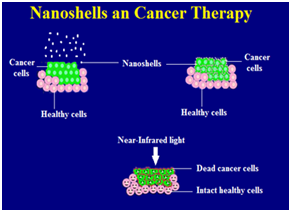
Fig: 7 nanoshells in cancer therapy
5. Dendrimers:
* It is a man-made molecule.
* This shape gives them vast amounts of surface area.
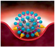
Fig: 8 dendrimer
A single dendrimer can carry a molecule that
* Recognizes Cancer Cells.
* A Therapeutic Agent To Kill Those Cells .
* A Molecule That Recognizes the Signals Of Cell Death.
NOW YOU CAN ALSO PUBLISH YOUR ARTICLE ONLINE.
SUBMIT YOUR ARTICLE/PROJECT AT articles@pharmatutor.org
Subscribe to Pharmatutor Alerts by Email
FIND OUT MORE ARTICLES AT OUR DATABASE
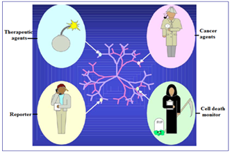
Fig: 9 molecules carrying by dendrimers
6. Cantilever:
* Coated with molecules (like antibodies).
* Capable of binding to specific molecules that only cancer cells secrete.
* Used in cancer detection.
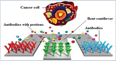
Fig: 10 nanomedicine-cantilever
Conclusion:
* Unique properties of cancer cells that can be exploited by nanoparticles to target the Cancer cells.
* On this basis highly specific nanoparticle production can be started for the discovery, treatment and prevention of cancer growth. It is safer and more consistent.
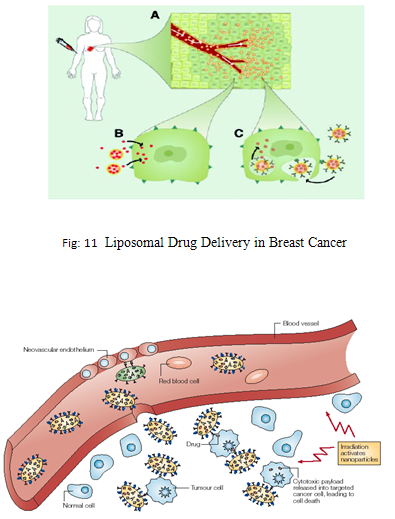
Fig: 12 Nanomedicine targeting the tumor cell
Visualization:
Tracking movement can help determine how well drugs are being distributed or how substances are metabolized. It is difficult to track a small group of cells throughout the body, so scientists used to dye the cells. These dyes needed to be excited by light of a certain wavelength in order for them to light up. While different color dyes absorb different frequencies of light, there was a need for as many light sources as cells.
A way around this problem is with luminescent tags. These tags are quantum dots attached to proteins that penetrate cell membranes. The dots can be random in size, can be made of bio-inert material, and they demonstrate the nanoscale property that color is size-dependent. As a result, sizes are selected so that the frequency of light used to make a group of quantum dots fluoresce is an even multiple of the frequency required to make another group incandesce. Then both groups can be lit with a single light source.
Nano Robots:
The somewhat speculative claims about the possibility of using nanorobots in medicine, advocates say, would totally change the world of medicineonce it is realized. Nanomedicine would make use of these nanorobots introduced into the body, to repair or detect damages and infections.
According to Robert Freitas of the Institute for Molecular Manufacturing, a typical blood borne medical nanorobot would be between 0.5-3 micrometres in size, because that is the maximum size possible due to capillary passage requirement. Carbon could be the primary element used to build these nanorobots due to the inherent strength and other characteristics of some forms of carbon (diamond/fullerenecomposites), and nanorobots would be fabricated in desktop nanofactories specialized for this purpose.
Nanodevices could be observed at work inside the body using MRI, especially if their components are isotope of carbon, since has a nonzero nuclear magnetic moment. Medical nanodevices would first be injected into a human body and would then go to work in a specific organ or tissue mass and make certain that the nanodevices have to get the correct target treatment region. The doctor will also be able to scan a section of the body, and actually see the nanodevices congregated neatly around their target, so that he or she can be sure that the procedure was successful.
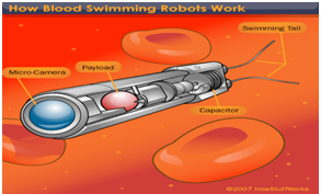
Fig: 13 nano robot
Cell Repair Machines:
Using drugs and surgery, doctors can only encourage tissues to repair themselves. With molecular machines, there will be more direct repairs cell repair will utilize the same tasks that living systems already prove possible. Access to cells is possible because biologists can insert needles into cells without killing them. Thus, molecular machines are capable of entering the cell. All specific biochemical interactions show that molecular systems can recognize other molecules by touch, build or rebuild every molecule in a cell, and can disassemble damaged molecules.
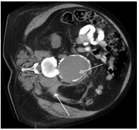
Fig: 14 implantation of nano medicine
Finally cells that replicate prove that molecular systems can assemble every system found in a cell. Therefore, since nature has demonstrated the basic operations needed to perform molecular-level cell repair, in the future, nanomachine based systems will be built that are able to enter cells, sense differences from healthy ones and make modifications to the structure.
The healthcare possibilities of these cell repair machines are impressive. Comparable to the size of viruses or bacteria, their compact parts would allow them to be more complex. The early machines will be specialized. As they open and close cell membranes or travel through tissue and enter cells and viruses, machines will only be able to correct a single molecular disorder like DNA damage or enzyme deficiency. Later, cell repair machines will be programmed with more abilities with the help of advanced AI systems.
Nano computers will be needed to guide these machines. These computers will direct machines to examine, take apart, and rebuild damaged molecular structures. Repair machines will be able to repair whole cells by working structure by structure. Then by working cell by cell and tissue by tissue, whole organs can be repaired. Finally, by working organ by organ, health is restored to the body. Cells damaged to the point of inactivity can be repaired because of the ability of molecular machines to build cells from scratch. Therefore, cell repair machines will free medicine from reliance on self repair alone.
Tissue Engineering:
Natural bone surface is quite often contains features that are about 100 nm across. If the surface of an artificial bone implant were left smooth, the body would try to reject it. Because of that smooth surface is likely to cause production of a fibrous tissue covering the surface of the implant. This layer reduces the bone-implant contact, which may result in loosening of the implant and further inflammation. It was demonstrated that by creating nano-sized features on the surface of the hip or knee prosthesis one could reduce the chances of rejection as well as to stimulate the production of osteoblasts. The osteoblasts are the cells responsible for the growth of the bone matrix and are found on the advancing surface of the developing bone.
The effect was demonstrated with polymeric, ceramic and, more recently, metal materials. More than 90% of the human bone cells from suspension adhered to the nanostructured metal surface, but only 50% in the control sample. In the end this findings would allow to design a more durable and longer lasting hip or knee replacements and to reduce the chances of the implant getting loose. Titanium is a well-known bone repairing material widely used in orthopaedics and dentistry. It has a high fracture resistance, ductility and weight to strength ratio. Unfortunately, it suffers from the lack of bioactivity, as it does not support cell adhesion and growth well. Apatite coatings are known to be bioactive and to bond to the bone. Hence, several techniques were used in the past to produce an appetite coating on titanium. Those coatings suffer from thickness non-uniformity, poor adhesion and low mechanical strength. In addition, a stable porous structure is required to support the nutrients transport through the cell growth.
It was shown that using a biomimetic approach – a slow growth of nanostructured apatite film from the simulated body fluid – resulted in the formation of a strongly adherent, uniform nanoporous layer. The layer was found to be built of 60 nm crystallites, and possess a stable nanoporous structure and bioactivity.A real bone is a nanocomposite material, composed of hydroxyapatite crystallites in the organic matrix, which is mainly composed of collagen.
An artificial hybrid material was prepared from 15–18 nm ceramic nanoparticles and poly (methyl methacrylate) copolymer. Using tribology approach, a viscoelastic behaviour (healing) of the human teeth was demonstrated. An investigated hybrid material, deposited as a coating on the tooth surface, improved scratch resistance as well as possessed a healing behaviour similar to that of the tooth.
Contact lens:
Researchers invent drug-dispensing contact lenses for the treatment of eye diseases. Nanoengineers in Singapore have invented a contact lens that can release precise amounts of medication to treat glaucoma and other eye diseases.
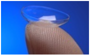
Fig: 15 nano medication in contact lens
Multicolor Optical Coding For Biological Assays:
The ever increasing research in proteomics and genomic generates escalating number of sequence data and requires development of high throughput screening technologies. Realistically, various array technologies that are currently used in parallel analysis are likely to reach saturation when a number of array elements exceed several millions. A three-dimensional approach, based on optical "bar coding" of polymer particles in solution, is limited only by the number of unique tags one can reliably produce and detect.
Single quantum dots of compound semiconductors were successfully used as a replacement of organic dyes in various bio-tagging applications. This idea has been taken one step further by combining differently sized and hence having different fluorescent colours quantum dots, and combining them in polymeric microbeads .A precise control of quantum dot ratios has been achieved. The selection of nanoparticles used in those experiments had 6 different colours as well as 10 intensities. It is enough to encode over 1 million combinations. The uniformity and reproducibility of beads was high letting for the bead identification accuracies of 99.99%.
Manipulation of cells and biomolecules:
Functionalized magnetic Nanoparticles have found many applications including cell separation and probing; these and other applications are discussed in a recent review . Most of the magnetic particles studied so far are spherical, which somewhat limits the possibilities to make these nanoparticles multifunctional. Alternative cylindrically shaped nanoparticles can be created by employing metal electrodeposition into nanoporous alumina template Depending on the properties of the template, nanocylinder radius can be selected in the range of 5 to 500 nm while their length can be as big as 60 μm.
As surface chemistry for functionalisation of metal surfaces is well developed, different ligands can be selectively attached to different segments. For example, porphyrins with thiol or carboxyl linkers were simultaneously attached to the gold or nickel segments respectively. Thus, it is possible to produce magnetic nanowires with spatially segregated fluorescent parts. In addition, because of the large aspect ratios, the residual magnetisation of these nanowires can be high. Hence, weaker magnetic field can be used to drive them. It has been shown that a self-assembly of magnetic nanowires in suspension can be controlled by weak external magnetic fields. This would potentially allow controlling cell assembly in different shapes and forms.
NOW YOU CAN ALSO PUBLISH YOUR ARTICLE ONLINE.
SUBMIT YOUR ARTICLE/PROJECT AT articles@pharmatutor.org
Subscribe to Pharmatutor Alerts by Email
FIND OUT MORE ARTICLES AT OUR DATABASE
Protein Detection:
Proteins are the important part of the cell's language, machinery and structure, and understanding their functionalities is extremely important for further progress in human well being. Gold nanoparticles are widely used in immunohistochemistry to identify protein-protein interaction. However, the multiple simultaneous detection capabilities of this technique are fairly limited. Surface-enhanced Raman scattering spectroscopy is a well-established technique for detection and identification of single dye molecules. By combining both methods in a single nanoparticle probe one can drastically improve the multiplexing capabilities of protein probes. The group of Prof. Mirkin has designed a sophisticated multifunctional probe that is built around a 13 nm gold nanoparticle.
The nanoparticles are coated with hydrophilic oligonucleotides containing a Raman dye at one end and terminally capped with a small molecule recognition element (e.g. biotin). Moreover, this molecule is catalytically active and will be coated with silver in the solution of Ag (I) and hydroquinone. After the probe is attached to a small molecule or an antigen it is designed to detect, the substrate is exposed to silver and hydroquinone solution. A silver-plating is happening close to the Raman dye, which allows for dye signature detection with a standard Raman microscope. Apart from being able to recognise small molecules this probe can be modified to contain antibodies on the surface to recognise proteins. When tested in the protein array format against both small molecules and proteins, the probe has shown no cross-reactivity.
Commercial Exploration:
The majority of the companies are small recent spinouts of various research institutions. Although not exhausting, this is a representative selection reflecting current industrial trends. Most of the companies are developing pharmaceutical applications, mainly for drug delivery. Several companies exploit quantum size effects in semiconductor nanocrystals for tagging biomolecules, or use bio-conjugated gold nanoparticles for labelling various cellular parts. A number of companies are applying nano-ceramic materials to tissue engineering and orthopaedics.
Most major and established pharmaceutical companies have internal research programs on drug delivery that are on formulations or dispersions containing components down to nano sizes. Colloidal silver is widely used in anti-microbial formulations and dressings. The high reactivity of titania nanoparticles, either on their own or then illuminated with UV light, is also used for bactericidal purposes in filters. Enhanced catalytic properties of surfaces of nano-ceramics or those of noble metals like platinum are used to destruct dangerous toxins and other hazardous organic materials.
As it stands now, the majority of commercial Nanoparticles applications in medicine are geared towards drug delivery. In biosciences, Nanoparticles are replacing organic dyes in the applications that require high photo-stability as well as high multiplexing capabilities. There are some developments in directing and remotely controlling the functions of Nano-probes, for example driving magnetic Nanoparticles to the tumor and then making them either to release the drug load or just heating them in order to destroy the surrounding tissue. The major trend in further development of nanomaterial is to make them multifunctional and controllable by external signals or by local environment thus essentially turning them into Nano-devices.
Nano nephrology:
Nanonephrology is a branch of nanomedicine and nanotechnology that seeks to use nano-materials and nano-devices for the diagnosis, therapy, and management of renal diseases. It includes the following goals:
· The study of kidney protein structures at the atomic level
· nano-imaging approaches to study cellular processes in kidney cells
· nano medical treatments that utilize nanoparticles to treat various kidney diseases
Advances in Nanonephrology are expected to be based on discoveries in the above areas that can provide nano-scale information on the cellular molecular machinery involved in normal kidney processes and in pathological states. By understanding the physical and chemical properties of proteins and other macromolecules at the atomic level in various cells in the kidney, novel therapeutic approaches can be designed to combat major renal diseases. The nano-scale artificial kidney is a goal that many physicians dream of. Nano-scale engineering advances will permit programmable and controllable nano-scale robots to execute curative and reconstructive procedures in the human kidney at the cellular and molecular levels. Designing nanostructures compatible with the kidney cells and that can safely operate in vivo is also a future goal. The ability to direct events in a controlled fashion at the cellular nano-level has the potential of significantly improving the lives of patients with kidney diseases.
Implants and Prosthetics:
The first field is implants and prosthetics. With the advent of new materials, and the synergy of nanotechnologies and biotechnologies, it could be possible to create artificial organs and implants that are more akin to the original, through cell growth on artificial scaffolds or biosynthetic coatings that increase biocompatibility and reduce rejection. These could include retinal, cochlear and neural implants, repair of damaged nerve cells, and replacements of damaged skin, tissue or bone.
Diagnostics Using Sensors and Micro Electro Mechanical Systems (MEMS):
The second area is diagnostics. Within MEMS (Micro Electro Mechanical Systems), laboratory-on-a-chip technology for quicker diagnosis which requires less of the sample is being developed in conjunction with microfluidics. In the medium term, it could be expected that general personal health monitors may be available. Developments in both genomics and nanotechnology are likely to enable sensors that can determine genetic make-up quickly and precisely, enhancing knowledge of people’s predisposition to genetic-related diseases.
Drug Delivery Using Nanoparticles and Molecular Carriers:
Finally, drug delivery is likely to benefit from the development of nanotechnology. With nanoparticles it is possible that drugs may be given better solubility, leading to better absorption. Also, drugs may be contained within a molecular carrier, either to protect them from stomach acids or to control the release of the drug to a specific targeted area, reducing the likelihood of side effects. Such drugs are already beginning pre-clinical or clinical trials, adhering to the strict regulatory requirements for new pharmaceuticals. Due to this, development costs are often high and outcomes of research sometimes limited.
Lab on a Chip and Advanced Drug Delivery Systems:
The ultimate combination of the laboratory-on-a-chip and advanced drug delivery technologies would be a device that was implantable in the body, which would continuously monitor the level of various biochemicals in the bloodstream and in response would release appropriate drugs. For example, an insulin-dependent diabetic could use such a device to continuously monitor and adjust insulin levels autonomously. There is no doubt that this is the direction that current advances in which microfluidics and drug delivery are heading.
Diagnostic and imaging techniques:
* Carbon nano tubes and gold nano particles are being used in a sensor that detects proteins indicative of oral cancer. Tests have shown this sensor to be accurate In detecting oral cancer and provides results in less than an hour.
* Silver nano rods in a diagnostic systems are being used to separate viruses, bacteria and other microscopic components of blood samples, allowing clearer Raman spectroscopy signals of the components. This methods have been demonstrated to allow identification of viruses, bacteria in less than an hour.
* Iron oxide nano particles can used to to improve MRI images of cancer tumors. The particles are coated with a peptides that binds to a cancer tumors, once the nano particles attached to the tumor the magnetic property of the iron oxide enhances the images from the magnetic resonance imagining scan.
* Anti microbial techniques:
* A nano particles cream has been shown to fight staph infections. The nano particles contain nitric oxide gas, which is known to kill bacteria.
* Studies on the mice have shown that using the nano particles cream to release nitric oxide gas at the site of staph abscesses significantly reduced the infection.
|
Company |
Product |
|
CytImmune |
Gold nanoparticles for targeted delivery of drugs to tumors |
|
NanoBio |
Nanoemulsions for nasal delivery to fight viruses (such as the flu and colds) or through the skin to fight bacteria |
|
NanoBioMagnetics |
Magnetically responsive nanoparticles for targeted drug delivery and other applications |
|
Nanosphere |
Diagnostic testing using gold nanoparticles to detect low levels of proteins indicating particular diseases |
|
Nanotherapeutics |
Nanoparticles for improving the performance of drug delivery by oral or nasal methods |
|
Z-Medica |
Medical gauze containing aluminosilicate nanoparticles which help blood clot faster in open wounds. |
|
Sirnaomics |
Nanoparticle enhanced techniques for delivery of siRNA |
|
Makefield Therapeutics |
Nanoparticle cream for delivery of nitric oxide gas to treat infection |
|
Traversa Therapeutics |
Delivery of siRNA molecules |
|
|
|
CONCLUSION
The application of nanotechnology to drug delivery has currently more than 20 Nanoparticles therapeutics are in clinical use, validating the ability of Nanoparticles to improve the therapeutic index of drugs. In addition to the already approved Nanoparticles, numerous other Nanoparticles platforms are currently under various stages of preclinical and clinical development, including various liposomes, polymeric micelles, dendrimers, quantum dots, gold Nanoparticles, and ceramic Nanoparticles. With continued research and development efforts, nanotechnology is expected to have a tremendous impact on medicine for decades to come.
Nanotechnology will change the very foundations of cancer diagnosis, treatment and prevention.The field of research that deals with the engineering and creation of things from materials that are less than 100 nanometers in size, especially single atoms or molecules. Nanotechnology is the creation of useful materials, devices, and systems through the manipulation of matter on this miniscule scale. The emerging field of nanotechnology involves scientists from many different disciplines, including physicists, chemists, engineers, and biologists. There are many interesting nanodevices being developed that have a potential to improve cancer detection, diagnosis, and treatment.
BIBLIOGRAPHY
1. Farokhzad, O.C. & Langer, R. Nanomedicine: developing smarter therapeutic and diagnostic modalities. Adv. Drug Deliv. Rev. 58, 1456–1459 (2006).
2. Liu, Y., Miyoshi, H. & Nakamura, M. Nanomedicine for drug delivery and imaging: a promising avenue for cancer therapy and diagnosis using targeted functional nanoparticles. Int. J. Cancer 120, 2527–2537(2007).
3. Brannon-Peppas, L. & Blanchette, J.O. Nanoparticle and targeted systems for cancer therapy. Adv. Drug Deliv. Rev. 56, 1649–1659 (2004).
4. Kawasaki, E.S. & Player, A. Nanotechnology, nanomedicine, and the development of new, effective therapies for cancer. Nanomedicine 1, 101–109 (2005).
5. Emerich, D.F. & Thanos, C.G. Targeted nanoparticle-based drug delivery and diagnosis. J. Drug Target. 15, 163–183 (2007).
6. Groneberg, D.A., Giersig, M., Welte, T. & Pison, U. Nanoparticle-based diagnosis and therapy. Curr. Drug Targets 7, 643–648 (2006).
7. Parak WJ, Gerion D, Pellegrino T, Zanchet D, Micheel C, Williams CS, Boudreau R, Le Gros MA, Larabell CA, Alivisatos AP: Biological applications of colloidal nanocrystals. Nanotechnology 2003, 14:R15-R27.
8. Pankhurst QA, Connolly J, Jones SK, Dobson J: Applications of magnetic nanoparticles in biomedicine. J Phys D: Appl Phys 2003, 36:R167-R181.
9. Ma J, Wong H, Kong LB, Peng KW: Biomimetic processing of nanocrystallite bioactive apatite coating on titanium. Nanotechnology 2003, 14:619-623.
10. De la Isla A, Brostow W, Bujard B, Estevez M, Rodriguez JR, Vargas S, Castano VM: Nanohybrid scratch resistant coating for teeth and bone viscoelasticity manifested in tribology. Mat Resr Innovat 2003, 7:110-114.
11. Gutwein LG, Webster TJ: Affects of alumina and titania nanoparticulates on bone cell function. American Ceramic Society 26 th Annual Meeting Conference Proceedings 2003, in press.
12. Roy I, Ohulchanskyy TY, Pudavar HE, Bergey EJ, Oseroff AR, Morgan J, Dougherty TJ, Prasad PN: Ceramic-based nanoparticles entrapping water-insoluble photosensitizing anticancer drugs: a novel drug-carrier system for photodynamic therapy. J Am Chem Soc 2003, 125:7860-7865.
13. Han M, Gao X, Su JZ, Nie S: Quantum-dot-tagged microbeads for multiplexed optical coding of biomolecules. Nature Biotechnology 2001, 19:631-635.
14. Reich DH, Tanase M, Hultgren A, Bauer LA, Chen CS, Meyer GJ: Biological applications of multifunctional magnetic nanowires. J Appl Phys 2003, 93:7275-7280.
15. Davis, F.F. The origin of pegnology. Adv. Drug Deliv. Rev. 54, 457–458 (2002).
16. Harries, M., Ellis, P. & Harper, P. Nanoparticle albumin-bound paclitaxel for metastatic breast cancer. J. Clin. Oncol. 23, 7768–7771 (2005).
17. Woodle, M.C. Controlling liposome blood clearance by surfacegrafted polymers. Adv. Drug Deliv. Rev. 32, 139–152 (1998).
18. Sapra, P. & Allen, T.M. Internalizing antibodies are necessary for improved therapeutic efficacy of antibody-targeted liposomal drugs. Cancer Res. 62, 7190–7194 (2002).
19. Simoes, S., Moreira, J.N., Fonseca, C., Duzgunes, N. & de Lima, M.C. On the formulation of pH-sensitive liposomes with long circulation times. Adv. Drug Deliv. Rev. 56, 947–965 (2004).
20. Bissett, D. et al. Phase I and pharmacokinetic (PK) study of MAG-CPT (PNU 166148): a polymeric derivative of camptothecin (CPT). Br. J. Cancer 91, 50–55 (2004).
21. Hoekstra, R. et al. Phase I and pharmacologic study of PKI166, an epidermal growth factor receptor tyrosine kinase inhibitor, in patients with advanced solid malignancies. Clin. Cancer Res. 11, 6908–6915.
22. Tiwari, S.B. & Amiji, M.M. Improved oral delivery of paclitaxel following administration in nanoemulsion formulations. J. Nanosci. Nanotechnol. 6, 3215–3221 (2006).
23. McCarthy, T.D. et al. Dendrimers as drugs: discovery and preclinical and clinical development of dendrimer-based microbicides for HIV and STI prevention. Mol. Pharm. 2, 312 318 (2005).
NOW YOU CAN ALSO PUBLISH YOUR ARTICLE ONLINE.
SUBMIT YOUR ARTICLE/PROJECT AT articles@pharmatutor.org
Subscribe to Pharmatutor Alerts by Email
FIND OUT MORE ARTICLES AT OUR DATABASE


